Review
. 2015 Nov 12;34(22):2735-57.
doi: 10.15252/embj.201591881. Epub 2015 Oct 21.

Complement activation, regulation, and molecular basis for complement-related diseases

Affiliations
Review

Complement activation, regulation, and molecular basis for complement-related diseases

Goran Bajic et al. EMBO J. .

Abstract

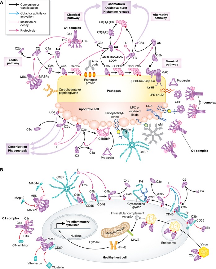
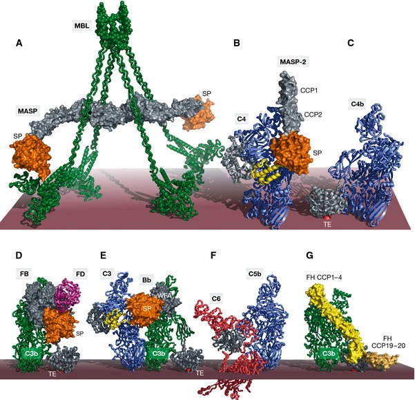
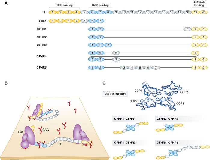
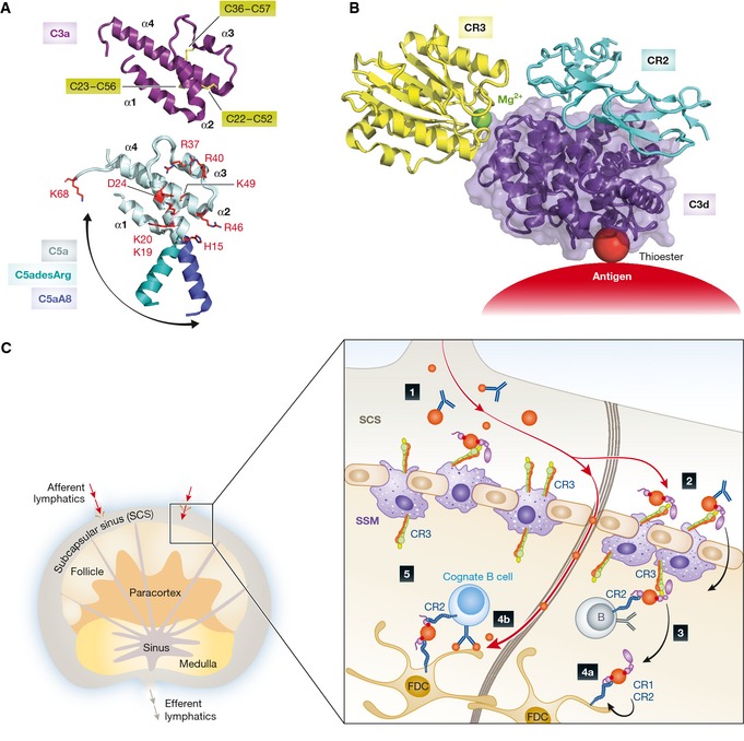
The complement system is an essential element of the innate immune response that becomes activated upon recognition of molecular patterns associated with microorganisms, abnormal host cells, and modified molecules in the extracellular environment. The resulting proteolytic cascade tags the complement activator for elimination and elicits a pro-inflammatory response leading to recruitment and activation of immune cells from both the innate and adaptive branches of the immune system. Through these activities, complement functions in the first line of defense against pathogens but also contributes significantly to the maintenance of homeostasis and prevention of autoimmunity. Activation of complement and the subsequent biological responses occur primarily in the extracellular environment. However, recent studies have demonstrated autocrine signaling by complement activation in intracellular vesicles, while the presence of a cytoplasmic receptor serves to detect complement-opsonized intracellular pathogens. Furthermore, breakthroughs in both functional and structural studies now make it possible to describe many of the intricate molecular mechanisms underlying complement activation and the subsequent downstream events, as well as its cross talk with, for example, signaling pathways, the coagulation system, and adaptive immunity. We present an integrated and updated view of complement based on structural and functional data and describe the new roles attributed to complement. Finally, we discuss how the structural and mechanistic understanding of the complement system rationalizes the genetic defects conferring uncontrolled activation or other undesirable effects of complement.

Keywords: complement; inflammation; innate immunity; proteolytic regulation; structural biology.


Figures

Figure 1
Figure 1. Molecular view of complement activation, amplification, and regulation
(A) Pattern recognition molecules sense the presence of pathogens and altered self. In the classical pathway, C1q (within the C1 complex) recognizes PAMPs (pathogen‐specific proteins, lipopolysaccharide (LPS), lipoteichoic acid (LTA), peptidoglycan) or DAMPs (DNA, phosphatidylserine, oxidized lipids, lysophosphatidylcholine (LPC)) either directly or antibody‐bound. This recognition induces autoactivation of C1r, which subsequently activates C1s. This is followed by cleavage of C4 and C2 by C1s and the subsequent formation of the CP C3 convertase C4b2a. Cleavage of C4 exposes an internal thioester, which causes C4b to become covalently attached to the activator surface, in turn tethering the convertase activity to the activator. In the lectin pathway, patterns of glycans are detected via MBL, CLLK, or ficolins leading to activation of MASPs and formation of the same C3 convertase, C4b2a. C3 convertases cleave C3 into C3b, which also becomes covalently attached to the activator surface. Surface‐associated C3b recruits FB, which leads to FB activation and the formation of C3bBb, the AP C3 convertase, which cleaves more C3 and amplifies complement activation. In addition to the surface‐bound C3 convertase, a fluid‐phase convertase can be formed by association of water‐reacted C3, termed C3(H20), to FB thus constantly maintaining a low level of complement activation in solution (tick‐over). Both of the surface‐bound C3 convertases can bind a C3b molecule whereby the C5 convertases are formed. These cleave C5 into C5a and C5b, thus initiating the terminal pathway and leading to formation of the membrane attack complex (MAC). Complement opsonins and PRMs are shown in purple, whereas the proteolytically active complexes are shown in light pink. (B) Complement activation and amplification are attenuated on host surfaces. The healthy cells express membrane‐bound or attract soluble regulators that irreversibly dissociate convertases (DAF, CR1, FH, C4BP) and act as cofactors for FI‐mediated degradation of C3b and C4b (MCP, FH, CR1, C4BP) or prevent MAC assembly (CD59). Soluble regulators also prevent formation of the MAC (clusterin, vitronectin). Recently, it was discovered that complement mediates a potent intracellular immune response to non‐enveloped viruses. Deposition and covalent attachment of C3 onto pathogens in the extracellular environment serve as a marker of cellular invasion because C3 products in the cytosol are detected by an as yet unidentified receptor. This receptor signals through MAVS and induces an antiviral state by triggering the transcription of pro‐inflammatory cytokines. Intracellular complement immunity is independent of professional immune cells and is conserved in mammals.
Figure 2
Figure 2. Large macromolecular complexes of complement proteins assembled upon complement activation
The order of panels (A–F) reflects the order of appearance starting from activation in the LP and ending with MAC assembly in the TP. (A) SAXS model of the MBL:MASP‐1 complex with MBL (green) associated with a MASP‐1 homodimer with its serine protease domains (orange) protruding away from the MBL collagen stems in agreement with an intercomplex activation mechanism. (B) Crystal structure of the C4:MASP‐2 complex (RCSB ID 4FXG) with the substrate (C4, blue with the anaphylatoxin domain in yellow) making contacts at two distinct sites; the CCP domains (gray) and the SP domain (orange). (C) Crystal structure of C4b (RSCB ID 4XAM) with the TE domain colored in gray and the reactive thioester covalently bound to the membrane shown as a red sphere. (D) Crystal structure of the ternary C3bB:D complex (RSCB ID 2XWB). FB binds C3b (green, with the TE domain in gray) via its vWA and 3 CCP domains (gray). The SP domain (orange) is in the closed state. FD (magenta) is recruited to FB. (E) Structural model of the AP C3 convertase in complex with a C3 substrate (blue) generated by superimposing C3bBb stabilized with SCIN (RCSB ID 2WIN) and the C5:CVF complex (RCSB ID 3PVM). The anaphylatoxin moiety (yellow) is released upon cleavage. (F) Crystal structure of the C5b6 complex (RCSB ID 4A5W) revealing conformational rearrangements occurring upon C5 cleavage to C5b (blue), reminiscent of those observed in the C3/C4 to C3b/C4b conversion. (G) Structural model of FH binding to C3b (green) generated by superimposing C3b bound to FH CCP1–4 (light yellow, RCSB ID 2WII) and TE domain bound to FH CCP19–20 (dark yellow, RCSB ID 4ONT), CCP5–18 are not illustrated. FH also interacts with host glycans. The binding of FH prepares C3b for FI binding and cleavage. In all panels, the red surface approximates the activator such as the surface of an LPS layer on a pathogenic bacterium. Importantly, this is separated from the cell membrane; thus, panel (F) does not imply that C6 extends into the membrane.
Figure 3
Figure 3. Complement factor H family regulators
(A) Domain organization of complement factor H (FH). C3‐binding domains are highlighted in yellow and glycosaminoglycan (GAG)‐contacting domains in blue. Complement factor H‐like 1 (FHL1) and complement factor H‐related (CFHR) proteins are represented below according to their sequence similarity to FH. CFHRs share high sequence similarity with each other and with FH. All CFHRs contain domains homologous to the FH GAG‐ and TED‐binding domains but lack the domains homologous to the FH N‐terminal CCP1–4. (B) Models of FH recruited to non‐activating surfaces. FH may be recruited to the self‐surface‐bound C3b and establish bivalent contacts with one C3b molecule. Owing to its flexible structure, FH may bind two surface‐bound C3b molecules (or iC3b and C3d). (C) CFHRs form homo‐ and heterodimers. The N‐terminal CCP1–2 domains of CFHR1 crystallize as head‐to‐tail dimers (RSCB ID 3ZD2). Due to the high sequence identity between the CCP1–2 of CFHR1, 2, and 5, the three proteins are able to form homo‐ and heterodimers in the serum and thus modulate complement activation in a more complex manner.
Figure 4
Figure 4. Structural characterization of anaphylatoxins and the role of CR3 and CR2 in antigen trafficking in the lymph node
(A) Structure of C3a and C5a anaphylatoxins. The overall structure of human C3a (RCSB ID 4HW5) indicates a four‐helix bundle fold (α‐helices are indicated) stabilized by three disulfide bridges (yellow sticks, cysteine numbering is indicated). Underneath is shown a superimposition of the C5a moiety from the intact C5 (light blue, RSCB ID 3CU7) with C5asdesArg (teal, RSCB ID 3HQB), and the C5aA8 antagonist (dark blue, RSCB ID 4P39). The swing‐out motion of the α1‐helix is indicated with an arrow. C5a residues involved in C5aR1 binding are indicated in red sticks. (B) Atomic model of the CR3 (yellow) and CR2 (light blue) co‐ligation to C3d (purple) generated by superimposing the CR3 I domain:C3d (RCSB ID 4M76) and CR2 CCP1‐2:C3d (RSCB ID 3OED) complexes. A ternary complex between a C3 opsonized antigen, CR2, and CR3 could be of physiological relevance, see the next panel. (C) Complement‐dependent antigen transport, uptake, recycling, and presentation occur in the lymph node. 1) Immune complexes (IC) containing complement‐opsonized antigens drain with the afferent lymphatics into the subcapsular sinus (SCS). 2) Complement‐opsonized antigen is taken up by subcapsular sinus macrophages (SSM) via complement receptor 3 (CR3) and shunted across the subcapsular sinus floor. 3) The antigen is handed off to non‐cognate B cells via complement receptor 2 (CR2), which transport it into the follicle. 4a) Antigen is delivered to follicular dendritic cells (FDCs) via CR1 and CR2. FDCs are subsequently able to retain antigen for long periods of time in a recycling compartment. 4b) Low‐molecular‐weight antigen is delivered directly into the follicle through conduits. 5) Cognate B cells can probe antigen arrayed on the surface of FDCs, and BCR signaling is enhanced by co‐ligation of CR2 by IC‐associated complement fragments. The color coding is consistent with panel (B).
Figure 5
Figure 5. The molecular basis of complement‐associated disease
(A) Crystal structure of the zymogen mutant G666E (red sticks) of the MASP‐3 CCP1‐CCP2‐SP fragment (RCSB ID 4KKD). The SP domain polymorphisms associated with the 3MC syndrome (H497Y, C630R, G666E, and G687R) are indicated. (B) Zoom‐in on the active site of the zymogen mutant G666E of MASP‐3. E666 is making multiple polar contacts (dashed black lines) with the α‐turn of the active site restraining the peptide chain in a locked position that keeps the catalytic residues S664 and H497 too far apart. (C) Zoom‐in on the catalytic pocket of the MASP‐1 SP domain (RCSB ID 4IGD). The equivalent of MASP‐3 E666 is G648 in MASP‐1, that makes only a single hydrogen bond, which is insufficient to restrain the catalytic S646 away from the catalytic H490. (D) Crystal structure of FH CCP6–8 in complex with sulfated glycans (RSCB ID 2UWN). All 3 CCP domains interact with host glycans. The carbohydrate‐binding residues are colored in blue. The H402 risk variant is highlighted in green. (E) FH CCP19–20 in complex with host‐specific glycans (RSCB ID 4ONT). The FH residues in contact with the TE domain of C3b, iC3b, or C3d is shown in pink; glycan‐interacting residues are shown in blue; aHUS‐associated mutations are shown in yellow; and aHUS‐associated mutations overlapping with glycan‐binding residues are highlighted in green. (F) Mapping of the aHUS‐associated C3 polymorphisms on C3b responsible for decreased cofactor activity of FH (green dot), MCP (pink dot), and both FH and MCP (light blue dot). Mapped is also the FH interaction area (yellow) explaining why aHUS is triggered when the polymorphisms occur on this C3b interface. (G) The C3b interaction area (cyan) is mapped on the surface of Bb (orange). aHUS‐associated polymorphisms of FB are highlighted with yellow dots. (H) Crystal structure of the CVF:C5 complex (RCSB ID 3PVM). The CVF interaction area (mapped in green) on C5 (blue) overlaps with that of the C5 convertase. Eculizumab prevents C5 recognition by the C5 convertases and is used in treatment of PNH. The red dot indicates the position of C5 Arg885, a polymorphism associated with the lack of response to eculizumab treatment of certain PNH patients. The eculizumab epitope comprises the Arg885 and thus overlaps with the putative convertase binding area.

References

    1. Agarwal S, Ferreira VP, Cortes C, Pangburn MK, Rice PA, Ram S (2010) An evaluation of the role of properdin in alternative pathway activation on Neisseria meningitidis and Neisseria gonorrhoeae . J Immunol 185: 507–516 - PMC - PubMed
    1. Alcorlo M, Tortajada A, Rodriguez de Cordoba S, Llorca O (2013) Structural basis for the stabilization of the complement alternative pathway C3 convertase by properdin. Proc Natl Acad Sci USA 110: 13504–13509 - PMC - PubMed
    1. Aleshin AE, DiScipio RG, Stec B, Liddington RC (2012) Crystal structure of C5b‐6 suggests structural basis for priming assembly of the membrane attack complex. J Biol Chem 287: 19642–19652 - PMC - PubMed
    1. Alper CA, Boenisch T, Watson L (1972) Genetic polymorphism in human glycine‐rich beta‐glycoprotein. J Exp Med 135: 68–80 - PMC - PubMed
    1. Amara U, Rittirsch D, Flierl M, Bruckner U, Klos A, Gebhard F, Lambris JD, Huber‐Lang M (2008) Interaction between the coagulation and complement system. Adv Exp Med Biol 632: 71–79 - PMC - PubMed

Publication types