Outcomes and second-look arthroscopic evaluation after combined arthroscopic treatment of tibial plateau and tibial eminence avulsion fractures: a 5-year minimal follow-up
- PMID: 26490156
- PMCID: PMC4618521
- DOI: 10.1186/s12891-015-0769-x
Outcomes and second-look arthroscopic evaluation after combined arthroscopic treatment of tibial plateau and tibial eminence avulsion fractures: a 5-year minimal follow-up
Abstract
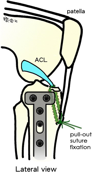
Background: Tibial eminence avulsion fracture often co-occurs with tibial plateau fracture, which leads to difficult concomitant management. The value of simultaneous arthroscopy-assisted treatment continues to be debated despite its theoretical advantages. We describe a simple arthroscopic suture fixation technique and hypothesize that simultaneous treatment is beneficial.
Methods: Patients with a tibial eminence avulsion fracture and a concurrent tibial plateau fracture who underwent simultaneous arthroscopically assisted treatment between 2005 and 2008 were enrolled in this retrospective study. Second-look arthroscopic evaluation and Rasmussen scores of clinical and radiographic parameters were used to assess simultaneous treatment.
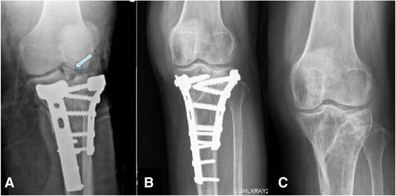
Results: Forty-one patients (41 knees) met the inclusion criteria. All 41 fractures were successfully united. All patients had side-to-side differences of less than 3 mm and negative findings in Lachman and pivot-shift tests at their final follow-up. The mean postoperative Rasmussen clinical score was 27.3 (range: 19-30), and the mean radiologic score was 16.5 (range: 12-18). Clinical and radiographic outcomes in 98 % of the patients were good or excellent. There were no complications directly associated with arthroscopy in any patient.
Conclusions: Simultaneous arthroscopic suture fixation of associated tibial eminence avulsion fracture did not interfere with the plates and screws used to stabilize the tibial plateau fracture. It gave the knee joint adequate stability, minimal surgical morbidity, and satisfactory radiographic and clinical outcomes in a minimum follow-up of 5 years and in the arthroscopic second-look assessments.
Figures



Similar articles
-
[Effectiveness of arthroscopic treatment of anterior cruciate ligament tibial eminence avulsion fracture with non-absorbable suture fixation combined with mini-plate].Zhongguo Xiu Fu Chong Jian Wai Ke Za Zhi. 2013 Sep;27(9):1041-4. Zhongguo Xiu Fu Chong Jian Wai Ke Za Zhi. 2013. PMID: 24279010 Chinese.
-
Comparative clinical efficacy of "Figure-8" Banding and double-row anchor suture-bridge fixation in arthroscopic management of tibial intercondylar eminence avulsion fractures.J Orthop Surg Res. 2024 Oct 16;19(1):663. doi: 10.1186/s13018-024-05111-1. J Orthop Surg Res. 2024. PMID: 39407246 Free PMC article.
-
[EFFECTIVENESS OF ARTHROSCOPIC ULTRA-Braid SUTURE PLANE FIXATION FOR ANTERIOR CRUCIATE LIGAMENT TIBIAL EMINENCE AVULSION FRACTURES].Zhongguo Xiu Fu Chong Jian Wai Ke Za Zhi. 2015 Nov;29(11):1353-7. Zhongguo Xiu Fu Chong Jian Wai Ke Za Zhi. 2015. PMID: 26875266 Chinese.
-
Arthroscopic-Assisted Reduction of Tibial Plateau Fractures.Orthop Clin North Am. 2019 Jul;50(3):305-314. doi: 10.1016/j.ocl.2019.03.011. Epub 2019 Apr 19. Orthop Clin North Am. 2019. PMID: 31084832 Review.
-
An original arthroscopic fixation of adult's tibial eminence fractures using the Tightrope® device: a report of 8 cases and review of literature.Knee. 2014 Aug;21(4):833-9. doi: 10.1016/j.knee.2014.02.007. Epub 2014 Feb 20. Knee. 2014. PMID: 24863950 Review.
Cited by
-
The efficacy of arthroscopy-assisted versus stand-alone open reduction and internal fixation for treating tibial plateau fracture: a systematic review and meta-analysis.BMC Musculoskelet Disord. 2024 Oct 30;25(1):865. doi: 10.1186/s12891-024-07958-1. BMC Musculoskelet Disord. 2024. PMID: 39472863 Free PMC article.
-
Tibial Eminence Avulsion in a Tibial Plateau Fracture - Our Approach: A Clinical Case.Rev Bras Ortop (Sao Paulo). 2021 Apr 19;59(2):e318-e322. doi: 10.1055/s-0041-1726067. eCollection 2024 Apr. Rev Bras Ortop (Sao Paulo). 2021. PMID: 38606129 Free PMC article.
-
The effectiveness of arthroscopy assisted fixation of Schatzker types I-III tibial plateau fractures: our experience at a tertiary centre.Int J Burns Trauma. 2021 Jun 15;11(3):163-169. eCollection 2021. Int J Burns Trauma. 2021. PMID: 34336380 Free PMC article.
-
Arthroscopic screw versus suture fixation in tibial eminence fractures: a systematic review and meta-analysis.Knee Surg Relat Res. 2025 Jul 24;37(1):31. doi: 10.1186/s43019-025-00282-5. Knee Surg Relat Res. 2025. PMID: 40708055 Free PMC article. Review.
References
-
- Wiley JJ, Baxter MP. Tibial spine fractures in children. Clin Orthop Relat Res. 1990;255:54–60. - PubMed
MeSH terms
LinkOut - more resources
Full Text Sources
Other Literature Sources
Medical
Miscellaneous

