. 2015 Oct 21:17:293.
doi: 10.1186/s13075-015-0811-2.

Analyzing pathogenic (double-stranded (ds) DNA-specific) plasma cells via immunofluorescence microscopy

Affiliations

Analyzing pathogenic (double-stranded (ds) DNA-specific) plasma cells via immunofluorescence microscopy

Oliver Winter et al. Arthritis Res Ther. .

Abstract

Introduction: While protective plasma cells (PCs) are an important part of the individual's immune defense, autoreactive plasma cells such as dsDNA-specific plasma cells contribute to the pathogenesis of autoimmune diseases like systemic lupus erythematosus (SLE). However, the research on dsDNA-specific plasma cells was restricted to the ELISpot technique, with its limitations, as no other attempt for identification of dsDNA-reactive plasma cells had been successful.

Methods: With improved fluorochrome labeling of dsDNA, removal of DNA aggregates, and enhanced blocking of unspecific binding, we were able to specifically detect dsDNA-reactive plasma cells by immunofluorescence microscopy.

Results: Via this novel technique we were able to distinguish short-lived (SLPCs) and long-lived (LLPCs) autoreactive plasma cells, discriminate dsDNA-specific plasma cells according to their immunoglobulin class (IgG, IgM, and IgA) and investigate autoreactive (dsDNA) and vaccine-induced ovalbumin (Ova) plasma cells in parallel.

Conclusions: The detection of autoreactive dsDNA-specific plasma cells via immunofluorescence microscopy allows specific studies on pathogenic and protective plasma cell subsets and their niches, detailed evaluation of therapeutic treatments and therefore offers new possibilities for basic and clinical research.


Figures

Fig. 1
Fig. 1
Analysis of double-stranded deoxyribonucleic acid (dsDNA)-specific plasma cells (PCs) by histology and ELISpot. a Kryosections from bone marrows and spleens of autoimmune (NZB/W) and non-autoimmune (C57BL/6) mice were stained with anti-immunoglobulin light chain kappa (IgL) (PCs, green) and fluorochrome-labeled dsDNA (red, - > dsDNA-specific PCs = yellow) and analyzed by confocal laser scanning microscopy. b For comparison standard enzymatic ELISpot (dsDNA of all isotypes) and immunofluorescence ELISpot (dsDNA red; IgG, IgA and IgM green) are depicted. For immunofluorescence ELISpot only 5 % of original cell numbers were seeded to enable counting of total PCs in parallel. c Numbers of dsDNA-specific PCs acquired by histology and ELISpot. With both methods, dsDNA PCs could be identified in autoimmune (NZB/W) but none to very few in non-autoimmune (C57BL/6) mice. d In probes from the same NZB/W mice, numbers of dsDNA-specific PCs were assessed by histology and ELISpot. Data in spleen and bone marrow correlated significantly
Fig. 2
Fig. 2
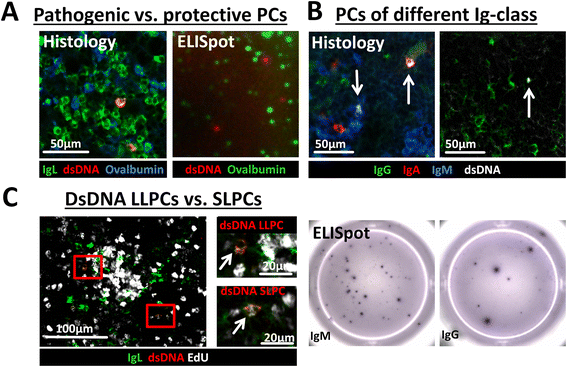
Parallel analysis of pathogenic (double-stranded deoxyribonucleic acid (dsDNA)-specific) and protective (ovalbumin (Ova)-specific) plasma cells (PCs), discrimination of dsDNA-specific PCs according to their immunoglobulin (Ig) class (IgA, IgG and IgM) and identification of autoreactive long-lived plasma cells (LLPCs) and short-lived plasma cells (SLPCs). a Kryosections from spleen of autoimmune mice (NZB/W) - 5 days post secondary Ova immunization - were stained with anti-Ig light chain kappa (IgL) (PCs, green), fluorochrome-labeled dsDNA (red, - > dsDNA-specific PCs = yellow) and fluorochrome-labeled Ova (blue, Ova-specific PCs = turquoise). PCs were either specific for dsDNA, Ova or an unknown antigen but no false double-positive dsDNA/Ova PCs occurred. For comparison, a figure of an immunofluorescence ELISpot identifying Ova (green) and dsDNA (red) antibody-secreting cells is depicted. b Kryosections from spleen of autoimmune (NZB/W) mice were stained with fluorochrome-labeled dsDNA (white), anti-IgA (red), anti-IgG (green), anti-IgM (blue). dsDNA-specific PCs were either IgM, IgG or IgA positive. The figures below depict dsDNA PCs of the IgM (left) and IgG (right) class acquired by ELISpot c Autoimmune (BcN/LmoJ) mice were fed EdU for 2 weeks. Kryosections from spleen were stained with click-it® EdU kit (EdU positive = white), IgL (PCs, green), fluorochrome-labeled dsDNA (red, - > dsDNA-specific PCs = yellow). dsDNA-specific SLPC incorporated EdU (lower selection, indicated by arrow in magnification, white-colored nucleus) during proliferation while dsDNA-specific LLPCs did not (upper selection, indicated by arrow in magnification, blank nucleus)

References

    1. Hiepe F, Dorner T, Hauser AE, Hoyer BF, Mei H, Radbruch A. Long-lived autoreactive plasma cells drive persistent autoimmune inflammation. Nat Rev Rheumatol. 2011;7:170–178. doi: 10.1038/nrrheum.2011.1. - DOI - PubMed
    1. Manz RA, Arce S, Cassese G, Hauser AE, Hiepe F, Radbruch A. Humoral immunity and long-lived plasma cells. Curr Opin Immunol. 2002;14:517–521. doi: 10.1016/S0952-7915(02)00356-4. - DOI - PubMed
    1. Naparstek Y, Plotz PH. The role of autoantibodies in autoimmune disease. Annu Rev Immunol. 1993;11:79–104. doi: 10.1146/annurev.iy.11.040193.000455. - DOI - PubMed
    1. Neves M, Alves JD. Factors implicated in the generation and persistence of long-lived plasma cell-mediated autoimmunity. Autoimmun Rev. 2011;10:375–382. doi: 10.1016/j.autrev.2010.12.007. - DOI - PubMed
    1. Wilson HL. B cells contribute to MS pathogenesis through antibody-dependent and antibody-independent mechanisms. Biologics. 2012;6:117–123. - PMC - PubMed

Publication types

MeSH terms