

Orbital Atherectomy Plaque Modification Assessment of the Femoropopliteal Artery Via Intravascular Ultrasound (TRUTH Study)
- PMID: 26490645
- PMCID: PMC4647186
- DOI: 10.1177/1538574415607361
Orbital Atherectomy Plaque Modification Assessment of the Femoropopliteal Artery Via Intravascular Ultrasound (TRUTH Study)
Abstract
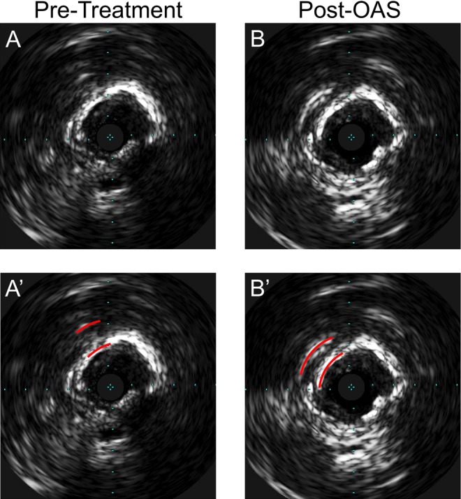
Objective: The Tissue Removal Assessment with Ultrasound of the SFA and Popliteal (TRUTH) study assessed the performance of the orbital atherectomy system (OAS) to treat femoropopliteal arteries, including determining its effect on plaque removal.
Methods: Patients with symptomatic femoropopliteal peripheral arterial disease were treated with the OAS followed by adjunctive balloon angioplasty (BA). Intravascular ultrasound (IVUS) images were collected pre- and post-OAS and post-OAS BA. Patients were followed through 12 months post-procedure.
Results: Twenty-nine lesions were treated with OAS-BA in 25 patients. The mean maximum balloon inflation pressure was 5.2 ± 1.2 atm. Virtual histology IVUS (VH-IVUS) analysis revealed at the maximum calcium ablation site that calcium reduction was responsible for 86% of the lumen area increase. The minimum lumen area increased from 4.0 mm(2) to 9.1 mm(2) (<.0001), and the percentage of area stenosis decreased from 76.9% to 43.0% (<.0001) after OAS-BA. At 12 months, the target lesion revascularization rate was 8.2%, and ankle-brachial index and Rutherford classification improved significantly from baseline through follow-up.
Conclusion: The VH-IVUS analysis reveals that OAS modifies the calcified component of the plaque burden. It is hypothesized that calcium modification by OAS changes the lesion compliance, allowing for low pressure adjunctive BA. The clinical outcomes were favorable through 12-month follow-up.
Trial registration: ClinicalTrials.gov NCT01938391.
Keywords: calcification; intravascular ultrasound; orbital atherectomy; peripheral arterial disease.
© The Author(s) 2015.
Conflict of interest statement
Figures


References
-
- Fitzgerald PJ, Ports TA, Yock PG. Contribution of localized calcium deposits to dissection after angioplasty. An observational study using intravascular ultrasound. Circulation. 1992;86(1):64–70. - PubMed
-
- Schwarzwälder U, Zeller T. Debulking procedures: potential device specific indications. Tech Vasc Interv Radiol. 2010;13(1):43–53. - PubMed
-
- Safian RD, Niazi K, Runyon JP, et al. Orbital atherectomy for infrapopliteal disease: device concept and outcome data for the OASIS trial. Catheter Cardiovasc Interv. 2009;73(3):406–412. - PubMed
-
- Adams GL, Khanna PK, Staniloae CS, Abraham JP, Sparrow EM. Optimal techniques with the Diamondback 360° System achieve effective results for the treatment of peripheral arterial disease. J Cardiovasc Transl Res. 2011;4(2):220–229. - PubMed
Publication types
MeSH terms
Associated data
LinkOut - more resources
Full Text Sources
Other Literature Sources
Medical
Research Materials
Miscellaneous
