. 2015 Oct 21:46:120.
doi: 10.1186/s13567-015-0265-1.

Infectious salmon anaemia virus (ISAV) mucosal infection in Atlantic salmon

Affiliations

Infectious salmon anaemia virus (ISAV) mucosal infection in Atlantic salmon

Maria Aamelfot et al. Vet Res. .

Abstract

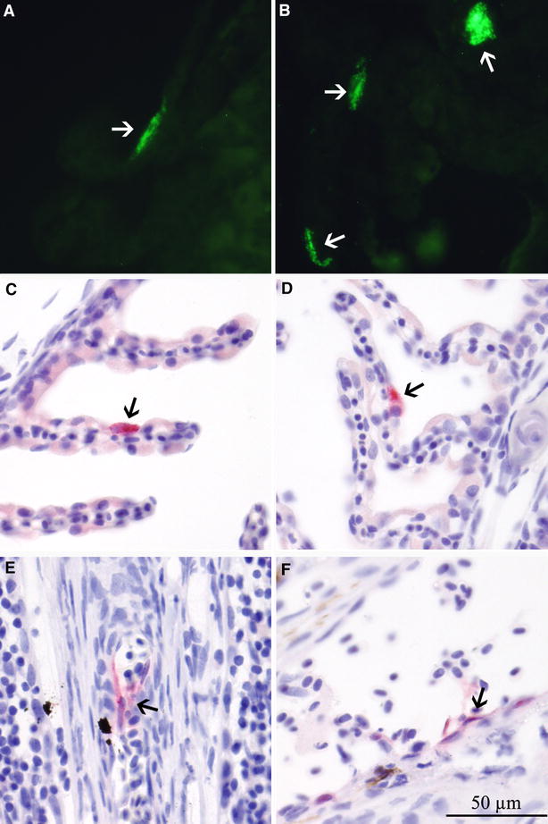
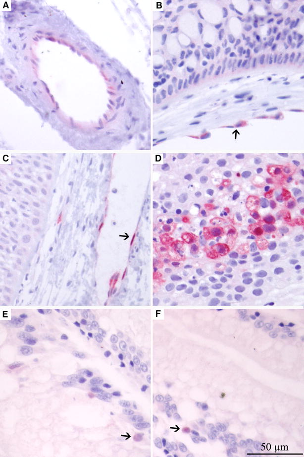
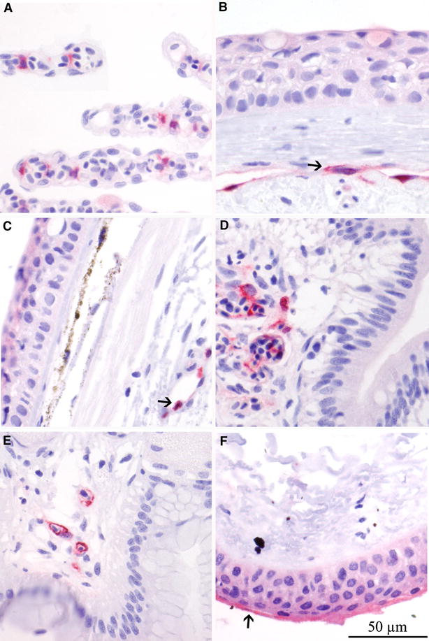
All viruses infecting fish must cross the surface mucosal barrier to successfully enter a host. Infectious salmon anaemia virus (ISAV), the causative agent of the economically important infectious salmon anaemia (ISA) in Atlantic salmon, Salmo salar L., has been shown to use the gills as its entry point. However, other entry ports have not been investigated despite the expression of virus receptors on the surface of epithelial cells in the skin, the gastrointestinal (GI) tract and the conjunctiva. Here we investigate the ISAV mucosal infection in Atlantic salmon after experimental immersion (bath) challenge and in farmed fish collected from a confirmed outbreak of ISA in Norway. We show for the first time evidence of early replication in several mucosal surfaces in addition to the gills, including the pectoral fin, skin and GI tract suggesting several potential entry points for the virus. Initially, the infection is localized and primarily infecting epithelial cells, however at later stages it becomes systemic, infecting the endothelial cells lining the circulatory system. Viruses of low and high virulence used in the challenge revealed possible variation in virus progression during infection at the mucosal surfaces.


Figures

Figure 1
Figure 1
ISAV segment 8 profiles measured by qPCR. Statistical modelling of ISAV segment 8 total RNA load profiles measured by real-time RT-qPCR of high virulent (dashed line and triangles) and low virulent (solid line and circles) strains from skin swabs (A), pectoral fin (B) and hind gut (C) post-immersion infection on day 0. Dotted lines indicate 95% confidence intervals.
Figure 2
Figure 2
IFAT and IHC of gills from challenged Atlantic salmon. IFAT (A, B) and IHC (CF) labelling of gill samples from challenged Atlantic salmon showing cells positive for ISAV HE and NP respectively. A, B Positive epithelial cells on gill lamellae, day 2 post infection (pi) LVI. C, D Positive epithelial cells on gill lamellae, day 5 pi LVI. E, F Positive endothelial cells in the gill, day 6 pi HVI. Arrows show positive cells.
Figure 3
Figure 3
Number of fish positive for ISAV HE in gill cryo-sections by IFAT. Fish positive on days 1–5 pi displayed positive epithelial cells, while fish positive on days 6–12 pi displayed positive endothelial cells. A transition from epithelial infection (days 2–5 pi) into endothelial infection (days 6–12 pi) occurs between day 5 and 6.
Figure 4
Figure 4
IHC labelling of pectoral fin/skin and gut from challenged Atlantic salmon. IHC labelling of pectoral fin/skin and gut samples from challenged Atlantic salmon showing cells positive for ISAV NP. A Positive endothelial cells (ECs) in vessel in skin, day 3 pi HVI. B, C Positive ECs in vessel in skin, day 4 pi HVI. D Skin of LVI on day 23 pi, showing necrotic epithelial cells in skin positive for ISAV. E, F Positive epithelial cells (EPs) in hind gut, day 4 pi LVI. Arrows show positive cells.
Figure 5
Figure 5
IHC labelling of mucosal tissues from Atlantic salmon from outbreak. IHC labelling of mucosal tissues from Atlantic salmon from confirmed outbreak of ISA showing cells positive for ISAV NP. Gill (A), pectoral fin (B), side-line skin (C), hind gut (D) and mid gut (E) with ISAV positive endothelial cells. Conjunctiva (F) with positive epithelial cells. Arrows show positive cells.
Figure 6
Figure 6
ISAV segment 8 measured by qPCR. Log of RT-qPCR ISAV segment 8 load in fish 1–5 from outbreak of ISA standardised to ELF and mean of 5 fish with standard error.

Similar articles

Cited by

References

    1. Dannevig BH, Falk K, Namork E. Isolation of the causal virus of infectious salmon anaemia (ISA) in a long-term cell line from Atlantic salmon head kidney. J Gen Virol. 1995;76:1353–1359. doi: 10.1099/0022-1317-76-6-1353. - DOI - PubMed
    1. Håstein T, Hill BJ, Winton JR. Successful aquatic animal disease emergency programmes. Rev Sci Tech. 1999;18:214–227. - PubMed
    1. Thorud K, Djupvik HO. Infectious anaemia in Atlantic salmon (Salmo salar L) Bull Eur Assoc Fish Pathol. 1988;8:109–111.
    1. Aamelfot M, Dale OB, Falk K. Infectious salmon anaemia—pathogenesis and tropism. J Fish Dis. 2014;37:291–307. doi: 10.1111/jfd.12225. - DOI - PubMed
    1. Roberts RJ. Fish pathology. London: W B Saunders; 2001.

Publication types

LinkOut - more resources