. 2015 Oct 21;35(42):14132-47.
doi: 10.1523/JNEUROSCI.0261-15.2015.

The Periaqueductal Gray Orchestrates Sensory and Motor Circuits at Multiple Levels of the Neuraxis

Affiliations

The Periaqueductal Gray Orchestrates Sensory and Motor Circuits at Multiple Levels of the Neuraxis

Stella Koutsikou et al. J Neurosci. .

Abstract

The periaqueductal gray (PAG) coordinates behaviors essential to survival, including striking changes in movement and posture (e.g., escape behaviors in response to noxious stimuli vs freezing in response to fear-evoking stimuli). However, the neural circuits underlying the expression of these behaviors remain poorly understood. We demonstrate in vivo in rats that activation of the ventrolateral PAG (vlPAG) affects motor systems at multiple levels of the neuraxis through the following: (1) differential control of spinal neurons that forward sensory information to the cerebellum via spino-olivo-cerebellar pathways (nociceptive signals are reduced while proprioceptive signals are enhanced); (2) alterations in cerebellar nuclear output as revealed by changes in expression of Fos-like immunoreactivity; and (3) regulation of spinal reflex circuits, as shown by an increase in α-motoneuron excitability. The capacity to coordinate sensory and motor functions is demonstrated in awake, behaving rats, in which natural activation of the vlPAG in fear-conditioned animals reduced transmission in spino-olivo-cerebellar pathways during periods of freezing that were associated with increased muscle tone and thus motor outflow. The increase in spinal motor reflex excitability and reduction in transmission of ascending sensory signals via spino-olivo-cerebellar pathways occurred simultaneously. We suggest that the interactions revealed in the present study between the vlPAG and sensorimotor circuits could form the neural substrate for survival behaviors associated with vlPAG activation.

Significance statement: Neural circuits that coordinate survival behaviors remain poorly understood. We demonstrate in rats that the periaqueductal gray (PAG) affects motor systems at the following multiple levels of the neuraxis: (1) through altering transmission in spino-olivary pathways that forward sensory signals to the cerebellum, reducing and enhancing transmission of nociceptive and proprioceptive information, respectively; (2) by alterations in cerebellar output; and (3) through enhancement of spinal motor reflex pathways. The sensory and motor effects occurred at the same time and were present in both anesthetized animals and behavioral experiments in which fear conditioning naturally activated the PAG. The results provide insights into the neural circuits that enable an animal to be ready and able to react to danger, thus assisting in survival.

Keywords: cerebellum; fear; nociception; periaqueductal grey; proprioception; spinal cord.


Figures

Figure 1.
Figure 1.
Characteristics of spino-olivary projection neurons. a, Typical single case example of antidromic testing, demonstrating the following: (i) the constant latency of the antidromically evoked spike (five consecutive trials superimposed), (ii) the ability to follow high-frequency stimulation (200 Hz), and (iii) collision (asterisk) with an orthodromically evoked spike. b, Mean ± SEM of the thresholds (n = 5) for antidromic activation as a function of the position of the stimulating electrode in the IO complex. Zero indicates the location of the stimulating electrode at a depth where the minimum current was required to evoke an antidromic spike. This location coincided stereotactically with the IO and was confirmed histologically. c, Distribution of the antidromic activation latencies of all spino-olivary neurons according to receptive field class, including neurons with unidentified peripheral receptive field (No RF). d, Histological identification of location of stimulation sites in the IO (two sites were not recovered) plotted on standard transverse maps of the IO. MAO, Medial accessory olive; PO, principal olive; DC, dorsal cap; VLO, ventrolateral outgrowth. Purple indicates class 1; green, class 2; red, class 3; and blue, class 4.
Figure 2.
Figure 2.
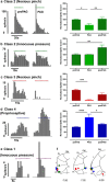
Ventrolateral PAG stimulation selectively alters responses to different qualities of sensory input of spino-olivary projection neurons. a, Typical example of the response of a class 2 neuron to noxious pinch (3.6 N): peristimulus time histogram (PSTH, spikes per 1 s bin) are shown before (pre-PAG) and during (PAG) vlPAG chemical excitation. Dotted horizontal line in each of the PSTHs indicates the onset and duration of the peripheral stimulus. Bar chart shows the average effect of vlPAG stimulation on all class 2 neuronal responses to noxious pinch (n = 7 neurons) before (pre-PAG), during (PAG), and after (post-PAG) microinjection of DLH into vlPAG. b, Same as a except example class 2 neuron response to innocuous pressure (0.5 N; n = 3 neurons). c, Same as a except example of class 3 neuron response to noxious pinch (3.6 N; n = 6 neurons). d, Same as a except example of class 4 neuron response to innocuous ankle joint manipulation (n = 8 neurons). All data are expressed as mean ± SEM of normalized spike counts in response to natural stimuli on the receptive field. *p < 0.05, **p < 0.01, ***p < 0.001, ****p < 0.0001 using repeated-measures ANOVA followed by Dunnett's post test versus pre-PAG. e, Example of the response of a single class 1 neuron to innocuous pressure (0.5 N): PSTH as described in a. f, Standard transverse maps of the left PAG at three rostrocaudal levels to show histological reconstruction of injection sites in all but three cases in which tissue could be recovered. In every case, the site of injection was verified physiologically with a transient drop in blood pressure in response to microinjection of DLH into vlPAG. Coordinates are relative to bregma. DM, Dorsomedial; DL, dorsolateral; L, lateral; VL, ventrolateral. Purple indicates class 1; green, class 2 (noxious pinch); green with black outline, class 2 (noxious pinch and innocuous pressure); red, class 3; blue, class 4.
Figure 3.
Figure 3.
Effects of noxious stimulation and vlPAG activation on FLI expression in the A and A2 subdivisions of the cerebellar nuclei. a, Standard transverse sections of the right hand cerebellar nuclei showing distribution of FLI neurons for four experimental groups, from left to right: microinjection of (i) saline into vlPAG (n = 7), (ii) microinjection of DLH into vlPAG (n = 7), (iii) anesthetic control (n = 8), and (iv) noxious pinch of the snout (n = 6). Each individual dot represents one FLI neuron. Results from all animals in each group are overlaid, b, Mean number of FLI neurons per section in the A and A2 subdivisions for animals in each experimental group. Data are represented as mean ± SEM. *p < 0.05, post hoc permutation t test with Bonferroni's correction. c, Standard transverse maps of the left PAG at two rostrocaudal levels to show histological reconstruction of injection sites of DLH (filled circles) and saline (open circles). no-STx, No stereotaxy involved in experiment (i.e., no nose clamp or ear bars were used), STx; stereotaxy involved in experiments (i.e., nose clamp and ear bars were used).
Figure 4.
Figure 4.
Effects of noxious stimulation and vlPAG activation on FLI expression in the AX subdivision of the cerebellar nuclei and control experiments with nitroprusside. a, Mean number of FLI neurons per section in the AX subdivision of the MCN for animals in each experimental group. No statistically significant differences were observed in the groups with microinjection of saline into vlPAG (n = 7), microinjection of DLH into vlPAG (n = 7), anesthetic control (n = 8), and noxious pinch of the snout (n = 6, p > 0.05, permutation one-way ANOVA). b, Mean number of FLI neurons per section in different subdivisions of MCN for anesthetic control (Anesth) and nitroprusside (Nitro) control groups. No significant differences in FLI in the MCN were observed between animals administered with sodium nitroprusside (n = 4) and anesthetic control animals (n = 8, p > 0.05, permutation one-way ANOVA).
Figure 5.
Figure 5.
Characterization of hindlimb evoked CFPs in awake rat. a, Superimposition of 3 consecutive field potentials evoked by electrical stimulation of the ipsilateral hindlimb (1.5× threshold) in an awake rat (stimulus onset indicated by filled arrowhead). b, Sagittal section of cerebellum showing electrode position (lesion indicated by filled arrowhead) from which recordings shown in a were made. c, Top two traces, Example average field potential waveforms (10 consecutive trials) recorded simultaneously from two positions shown in the sagittal section of the cerebellum. Bottom trace, Simultaneously recorded neck EMG. d, Stimulus–response curve for CFPs (red dashed line) and EMG (black dashed line) after ipsilateral hindlimb stimulation (n = 7 rats). Stimulus intensity is expressed as multiples of the threshold (T) required to evoke a detectable cerebellar response. e, Effect of paired hindlimb stimulation on the amplitude of the early (black dashed line) and late component (red dashed line) of evoked CFPs recorded in COP (n = 7 rats). f, Example CFP (top trace) and individual Purkinje cell complex spike (bottom trace) evoked by ipsilateral hindlimb stimulation (filled arrowhead) recorded from the same position in COP in one rat. COP, copula pyramidis; CI, crus I; CII, crus II; PML, paramedian lobule.
Figure 6.
Figure 6.
Evidence of modulation in olivocerebellar pathway transmission during freezing. a, In one animal, the excitatory amino acid DLH was injected (100 nl, 50 mm; dashed line indicates time of injection) into the vlPAG while recording CFP responses evoked by ipsilateral hindlimb stimulation. DLH injection resulted in a reduction in CFP amplitude, together with a robust expression of freezing-like behavior (horizontal black bar indicates period in which the rat spent 95% of time in freezing-like behavior; light gray bars indicate baseline (55%) and recovery (52%) levels of inactivity, respectively). b, Camera lucida drawing of transverse view of PAG (−8.16 mm relative to bregma) showing location of bilateral injection cannulae (indicated by filled areas). c, Group data from fear conditioning experiments in which the amplitude of evoked CFPs was measured during periods of spontaneous inactivity (open bar, before fear recall) and during identified freezing epochs (filled bar, after exposure to previously conditioned stimuli). ***p < 0.001, paired t test; n = 5 rats. d, EMG amplitude during the same conditions as in c (*p < 0.05, paired t test; n = 5 rats). dm, Dorsomedial PAG; lat, lateral PAG; dl, dorsolateral PAG.
Figure 7.
Figure 7.
Activation of vlPAG results in simultaneous, differential modulation of SOCP transmission and spinal reflex circuits. ai, Example CFPs recorded from the surface of the cerebellar cortex (C1 zone of left copula pyramidis). aii, Examples of averaged M-wave (M) and H-reflex (H) responses recorded from the left plantaris muscle at the same time as ai. All responses were evoked by electrical stimulation of the ipsilateral tibial nerve (<1 mA). Each example consists of five consecutive responses averaged before (pre-PAG) and during (PAG) vlPAG chemical excitation with DLH. Arrows indicate onset of the electrical stimulus. b, Group data (mean ± SEM) showing that microinjections of DLH into the vlPAG facilitate the mean H-reflex amplitude expressed relative to the size of the M-wave (H:M ratio) during (PAG) vlPAG neuronal activation (open bars; left y-axis; n = 5, **p = 0.0025, F(2,72) = 10.45, repeated-measures ANOVA followed by Dunnett's post test vs pre-PAG). Simultaneously, vlPAG excitation reduces CFP responses evoked by the same electrical stimulus (hatched bars; right y-axis; n = 5, ****p < 0.0001, F(2,72) = 92.46, repeated-measures ANOVA followed by Dunnett's post test vs pre-PAG). c, Standard transverse maps of the left PAG to show location of injection sites of DLH in the vlPAG (filled circles). Coordinates are relative to bregma. DM, Dorsomedial; DL, dorsolateral; L, lateral; VL, ventrolateral.

Similar articles

Cited by

References

    1. Ackerley R, Pardoe J, Apps R. A novel site of synaptic relay for climbing fibre pathways relaying signals from the motor cortex to the cerebellar cortical C1 zone. J Physiol. 2006;576:503–518. doi: 10.1113/jphysiol.2006.114215. - DOI - PMC - PubMed
    1. Apps R. Movement-related gating of climbing fibre input to cerebellar cortical zones. Prog Neurobiol. 1999;57:537–562. doi: 10.1016/S0301-0082(98)00068-9. - DOI - PubMed
    1. Apps R. Gating of climbing fibre input to cerebellar cortical zones. Prog Brain Res. 2000;124:201–211. doi: 10.1016/S0079-6123(00)24017-X. - DOI - PubMed
    1. Apps R, Lee S. Gating of transmission in climbing fibre paths to cerebellar cortical C1 and C3 zones in the rostral paramedian lobule during locomotion in the cat. J Physiol. 1999;516:875–883. doi: 10.1111/j.1469-7793.1999.0875u.x. - DOI - PMC - PubMed
    1. Apps R, Hartell NA, Armstrong DM. Step phase-related excitability changes in spino-olivocerebellar paths to the c1 and c3 zones in cat cerebellum. J Physiol. 1995;483:687–702. doi: 10.1113/jphysiol.1995.sp020614. - DOI - PMC - PubMed

Publication types

MeSH terms

Substances

LinkOut - more resources