. 2015 Oct 21;35(42):14160-71.
doi: 10.1523/JNEUROSCI.0596-15.2015.

Decoding Internally and Externally Driven Movement Plans

Affiliations

Decoding Internally and Externally Driven Movement Plans

Giacomo Ariani et al. J Neurosci. .

Abstract

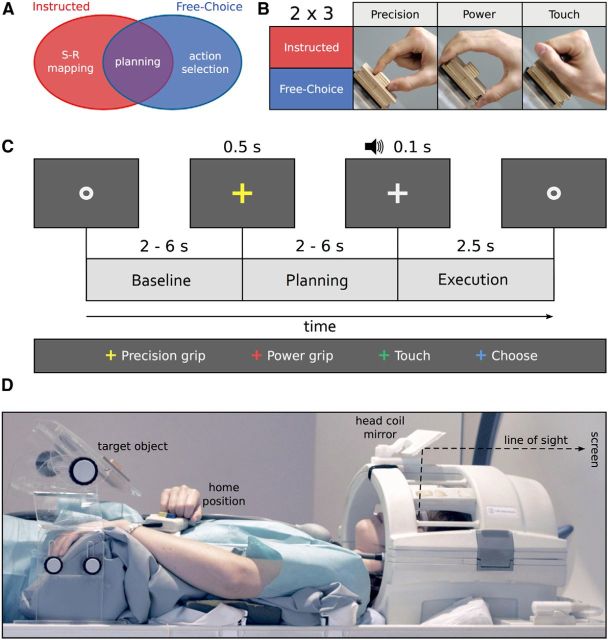
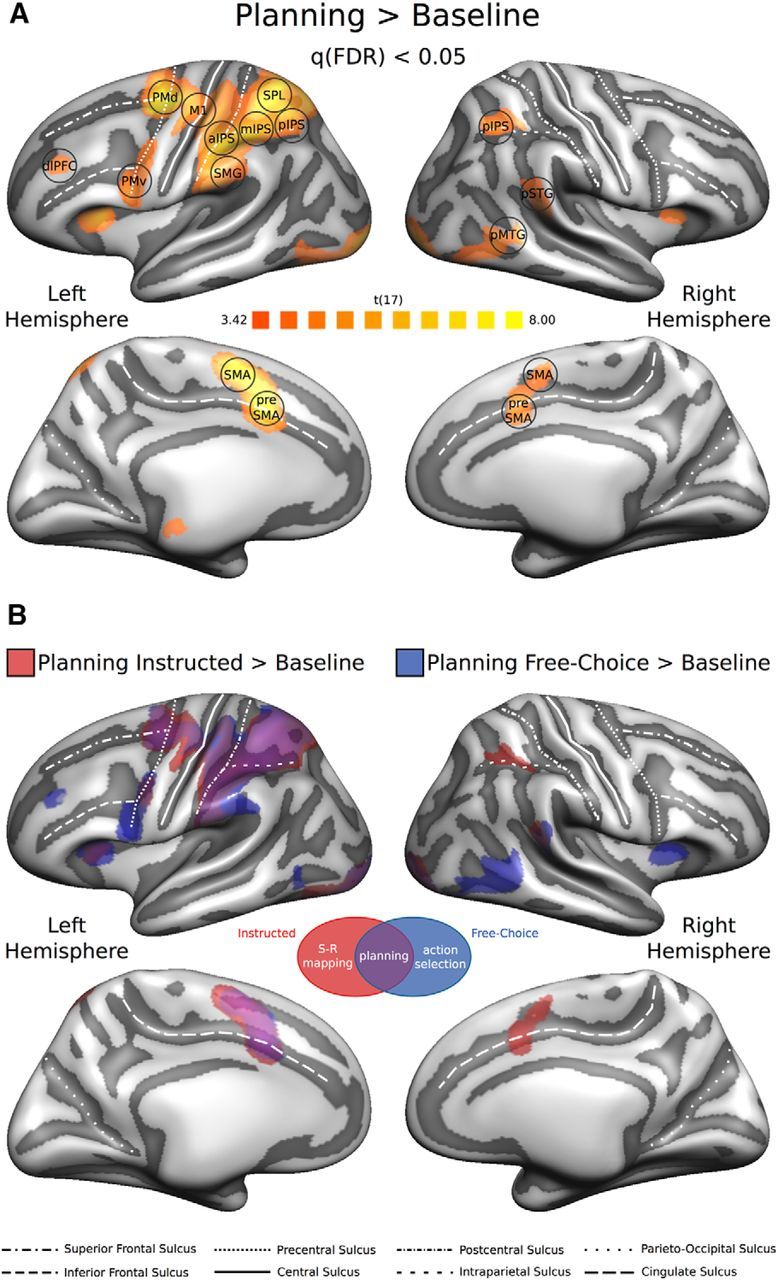
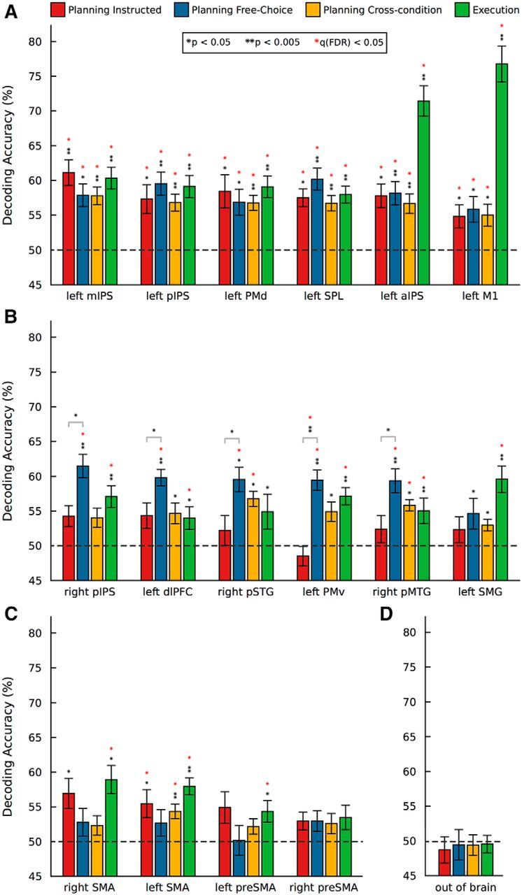
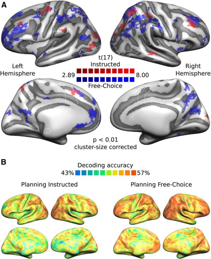
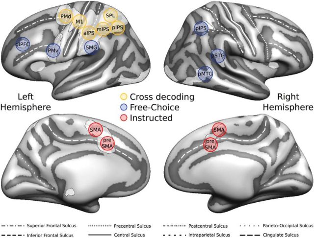
During movement planning, brain activity within parietofrontal networks encodes information about upcoming actions that can be driven either externally (e.g., by a sensory cue) or internally (i.e., by a choice/decision). Here we used multivariate pattern analysis (MVPA) of fMRI data to distinguish between areas that represent (1) abstract movement plans that generalize across the way in which these were driven, (2) internally driven movement plans, or (3) externally driven movement plans. In a delayed-movement paradigm, human volunteers were asked to plan and execute three types of nonvisually guided right-handed reaching movements toward a central target object: using a precision grip, a power grip, or touching the object without hand preshaping. On separate blocks of trials, movements were either instructed via color cues (Instructed condition), or chosen by the participant (Free-Choice condition). Using ROI-based and whole-brain searchlight-based MVPA, we found abstract representations of planned movements that generalize across the way these movements are selected (internally vs externally driven) in parietal cortex, dorsal premotor cortex, and primary motor cortex contralateral to the acting hand. In addition, we revealed representations specific for internally driven movement plans in contralateral ventral premotor cortex, dorsolateral prefrontal cortex, supramarginal gyrus, and in ipsilateral posterior parietotemporal regions, suggesting that these regions are recruited during movement selection. Finally, we observed representations of externally driven movement plans in bilateral supplementary motor cortex and a similar trend in presupplementary motor cortex, suggesting a role in stimulus-response mapping.

Significance statement: The way the human brain prepares the body for action constitutes an essential part of our ability to interact with our environment. Previous studies demonstrated that patterns of neuronal activity encode upcoming movements. Here we used multivariate pattern analysis of human fMRI data to distinguish between brain regions containing movement plans for instructed (externally driven) movements, areas involved in movement selection (internally driven), and areas containing abstract movement plans that are invariant to the way these were generated (i.e., that generalize across externally and internally driven movement plans). Our findings extend our understanding of the neural basis of movement planning and have the potential to contribute to the development of brain-controlled neural prosthetic devices.

Keywords: MVPA; decoding; fMRI; free choice; movement planning; movement selection.


Figures

Figure 1.
Figure 1.
Experimental question, design, timing, and setup. A, Schematic representation of the research question: is it possible to distinguish between areas representing externally triggered (instructed) movement plans (red), internally triggered (freely chosen) movement plans (blue), and abstract movement plans that are invariant to the way these movement plans are generated (purple)? B, A 2 × 3 mixed factorial design: Planning condition (Instructed, Free-Choice), blocked, and Movement type (precision grip, PRG: two fingers only, index and thumb; power grip, PWG: whole hand open; touch, TCH: hand closed in a fist, without hand preshaping), randomized. C, Example trial with timing (Instructed block, PRG). Each trial began with participants fixating a dot (Baseline) for a variable amount of time randomly selected from a geometric distribution (p = 0.3, 2000–6000 ms). This interval was followed by a color fixation cross (500 ms) either instructing which movement to plan (Instructed blocks), or indicating to freely select one of the movements (Free-Choice blocks). The Planning phase consisted of a jittered interstimulus interval (independently chosen from the same geometric distribution). After this delay, an auditory cue (100 ms) provided the GO-signal to start the movement (Execution phase, 2500 ms). In the Instructed condition, the color of the fixation cross corresponded to one of the three movements. In the Free-Choice condition, the cue always had the same, noninformative, color (in this example, blue). D, Lateral view of a participant with the right hand at the home position. The central wooden target object on which the reach-to-grasp movements were performed was mounted on a Plexiglas workspace positioned above the waist of the participant. The size of the small and large wooden cuboids were 2 × 2 × 1 and 7 × 7 × 2 cm, respectively. Participants saw the screen through a mirror attached to the head coil (line of sight illustrated by black dashed line). This setup ensured that participants saw neither the target object nor their own movements.
Figure 2.
Figure 2.
Univariate RFX-GLM analysis. A, The univariate contrast [Planning > Baseline] (collapsing across planning conditions) was used to identify ROIs preferentially involved in movement planning. The resulting statistical RFX group map (N = 18) was corrected for multiple comparisons using a FDR q < 0.05 and projected on the group-averaged inflated surface mesh for visualization. Individual ROIs were defined as spheres (8 mm radius) around individual peak voxels resulting from single-subject statistical maps. Black circles represent an example of the individual spherical ROIs (for additional details, see Materials and Methods; Table 1). B, Univariate contrast [Planning > Baseline], separately for each Planning condition (red represents [Planning Instructed > Baseline]; blue represents [Planning Free-Choice > Baseline]), projected on the same group-averaged inflated surface mesh. Purple areas represent the overlap between the two statistical group maps.
Figure 3.
Figure 3.
ROI-based MVPA. Mean percentage decoding accuracies for movement type resulting from multiple binary classifiers. SVM classification accuracies for the three possible discriminations between movement pairs were averaged to produce a unique score per ROI and planning condition. Red bars, Planning Instructed. Blue bars, Planning Free-Choice. Yellow bars, Planning cross-condition (see Materials and Methods). Green bars, Execution (collapsing across Planning conditions). Statistical significance was assessed via one-sample t tests (two-tailed) against 50% chance. Results were FDR-corrected for multiple comparisons (number of ROIs × number of tests). Significance levels: one black asterisk, uncorrected p < 0.05; two black asterisks, uncorrected p < 0.005; one red asterisk, FDR corrected q < 0.05. A, Regions where we found both significant within- and cross-condition decoding. B, Regions where we observed significant effects (or trends) for the Free-Choice, but not for the Instructed Planning task. C, Regions where we observed significant effects (or trends) for the Instructed, but not for the Free-Choice Planning task. D, Control non-brain region outside the brain.
Figure 4.
Figure 4.
Searchlight SVM-MVPA: cross-condition decoding. The spherical searchlight (8 mm radius) was restricted to the surface (−1 to 3 mm). Decoding procedures were very similar to the ones used for the ROI-based MVPA (see Materials and Methods). A, Group t map (thresholded at p < 0.01 and then cluster-size corrected) for the cross-condition decoding projected on the group-averaged surface mesh. White dashed lines indicate the outlines of the statistical map revealed by the univariate contrast [Planning > Baseline]. B, Group accuracy map (%) for cross-condition decoding.
Figure 5.
Figure 5.
Searchlight SVM-MVPA: within-condition decoding. A, Group t maps (thresholded at p < 0.01 and then cluster-size corrected), separately for each planning condition (red represents Instructed; blue represents Free-Choice), projected on the group-averaged surface mesh. B, Group decoding accuracy maps (%) separately for each planning condition (left, Planning Instructed; right, Planning Free-Choice). All other conventions are the same as in Figure 4.
Figure 6.
Figure 6.
Summary of decoding results for the Planning phase. Circles superimposed on the group-averaged surface mesh represent examples of individual spherical ROIs color-coded according to the results of the ROI MVPA: yellow represents significant cross-condition decoding; blue represents preferential decoding for Free-Choice planning; red represents preferential decoding for Instructed planning. White-shaded areas with dashed outlines represent the statistical map revealed by the univariate contrast [Planning > Baseline].

Similar articles

Cited by

References

    1. Afshar A, Santhanam G, Yu BM, Ryu SI, Sahani M, Shenoy KV. Single-trial neural correlates of arm movement preparation. Neuron. 2011;71:555–564. doi: 10.1016/j.neuron.2011.05.047. - DOI - PMC - PubMed
    1. Andersen RA, Buneo CA. Intentional maps in posterior parietal cortex. Annu Rev Neurosci. 2002;25:189–220. doi: 10.1146/annurev.neuro.25.112701.142922. - DOI - PubMed
    1. Andersen RA, Cui H. Intention, action planning, and decision making in parietal-frontal circuits. Neuron. 2009;63:568–583. doi: 10.1016/j.neuron.2009.08.028. - DOI - PubMed
    1. Astafiev SV, Stanley CM, Shulman GL, Corbetta M. Extrastriate body area in human occipital cortex responds to the performance of motor actions. Nat Neurosci. 2004;7:542–548. doi: 10.1038/nn1241. - DOI - PubMed
    1. Badre D, D'Esposito M. Is the rostro-caudal axis of the frontal lobe hierarchical? Nat Rev Neurosci. 2009;10:659–669. doi: 10.1038/nrn2667. - DOI - PMC - PubMed

Publication types

MeSH terms

LinkOut - more resources