. 2015 Oct 21;35(42):14353-69.
doi: 10.1523/JNEUROSCI.1910-15.2015.

Metabolic Connection of Inflammatory Pain: Pivotal Role of a Pyruvate Dehydrogenase Kinase-Pyruvate Dehydrogenase-Lactic Acid Axis

Affiliations

Metabolic Connection of Inflammatory Pain: Pivotal Role of a Pyruvate Dehydrogenase Kinase-Pyruvate Dehydrogenase-Lactic Acid Axis

Mithilesh Kumar Jha et al. J Neurosci. .

Abstract

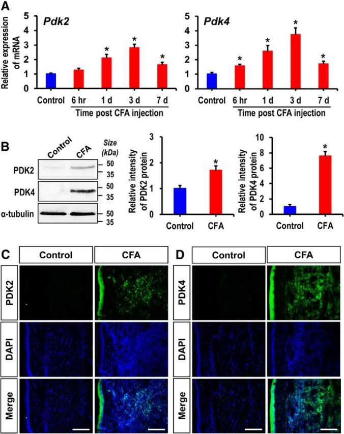
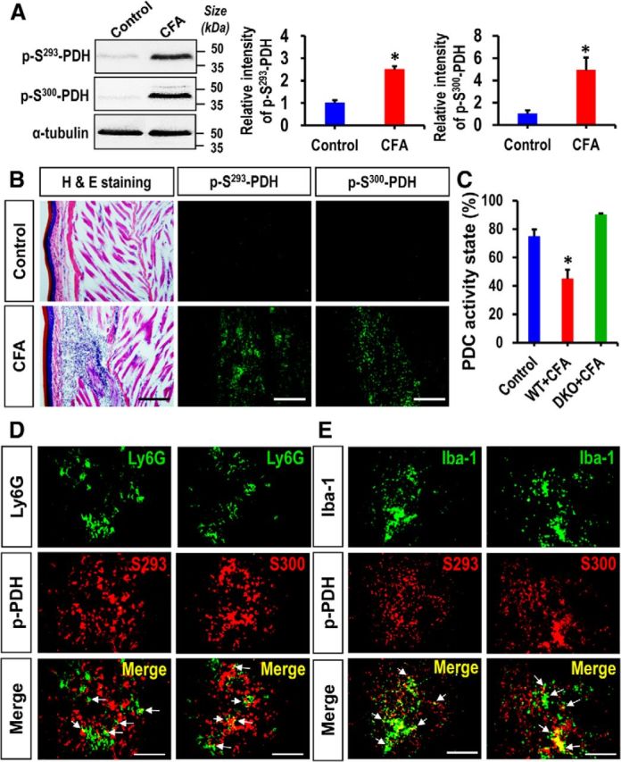
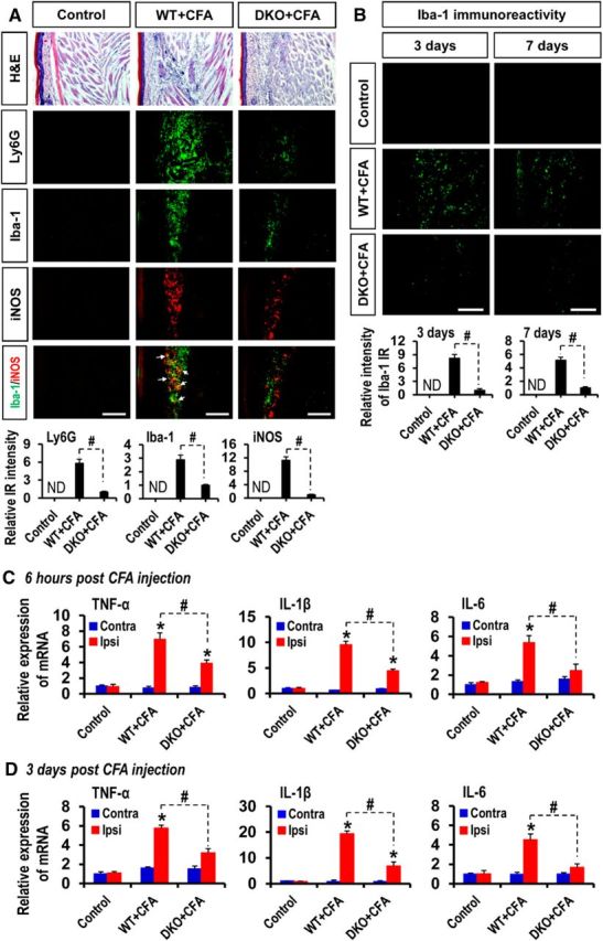
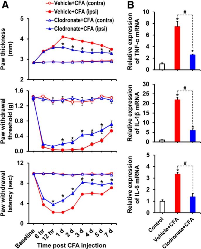
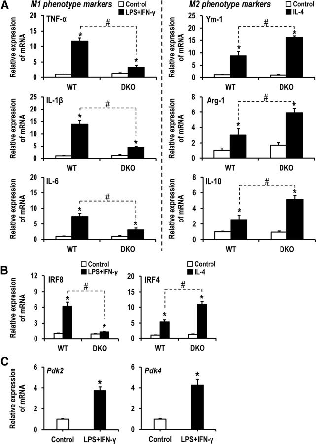
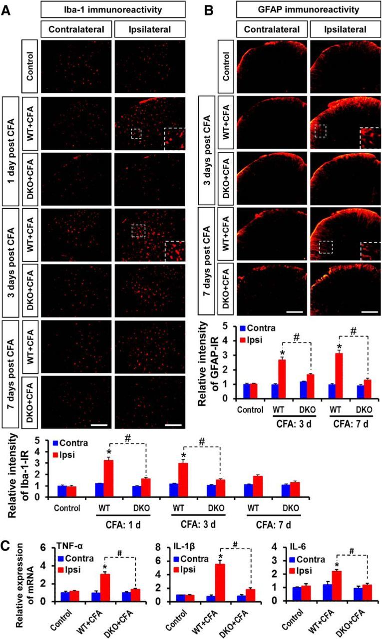
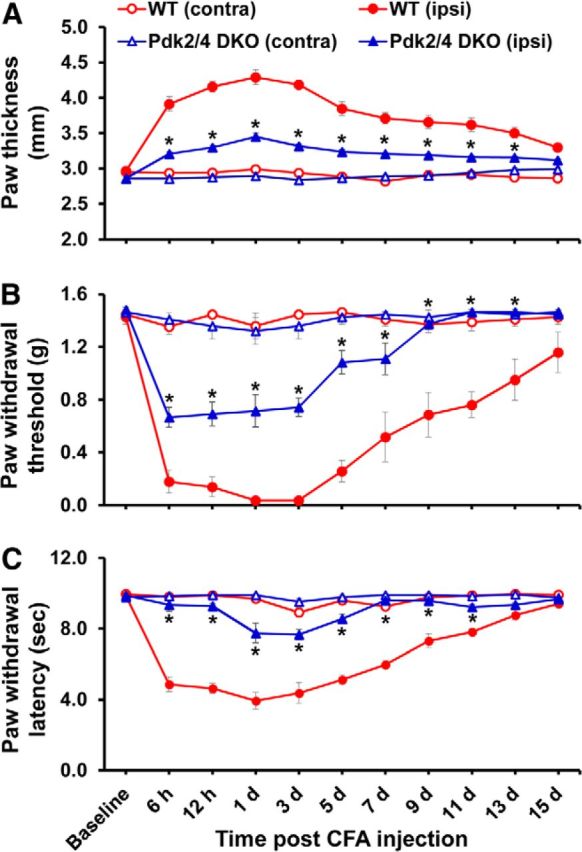
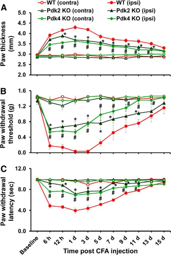
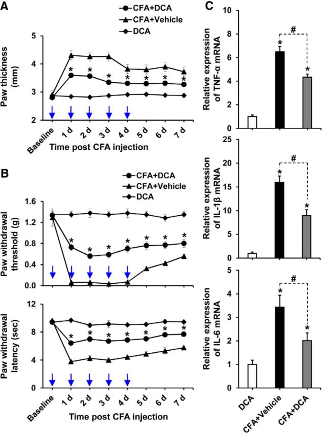
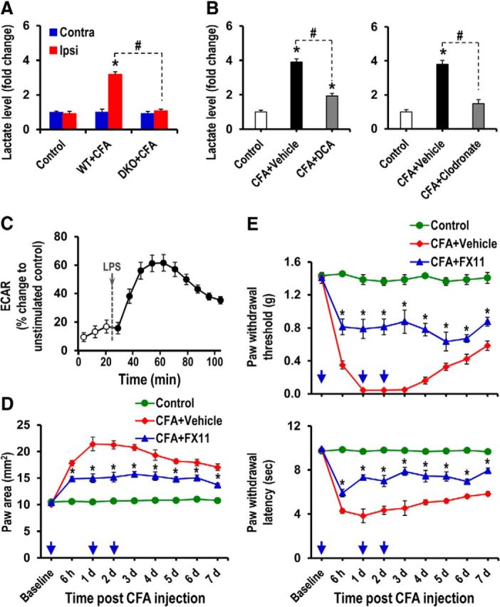
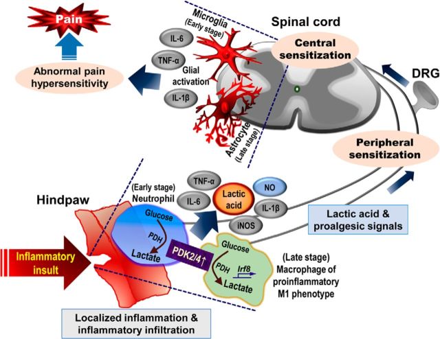
Pyruvate dehydrogenase kinases (PDK1-4) are mitochondrial metabolic regulators that serve as decision makers via modulation of pyruvate dehydrogenase (PDH) activity to convert pyruvate either aerobically to acetyl-CoA or anaerobically to lactate. Metabolic dysregulation and inflammatory processes are two sides of the same coin in several pathophysiological conditions. The lactic acid surge associated with the metabolic shift has been implicated in diverse painful states. In this study, we investigated the role of PDK-PDH-lactic acid axis in the pathogenesis of chronic inflammatory pain. Deficiency of Pdk2 and/or Pdk4 in mice attenuated complete Freund's adjuvant (CFA)-induced pain hypersensitivities. Likewise, Pdk2/4 deficiency attenuated the localized lactic acid surge along with hallmarks of peripheral and central inflammation following intraplantar administration of CFA. In vitro studies supported the role of PDK2/4 as promoters of classical proinflammatory activation of macrophages. Moreover, the pharmacological inhibition of PDKs or lactic acid production diminished CFA-induced inflammation and pain hypersensitivities. Thus, a PDK-PDH-lactic acid axis seems to mediate inflammation-driven chronic pain, establishing a connection between metabolism and inflammatory pain.

Significance statement: The mitochondrial pyruvate dehydrogenase (PDH) kinases (PDKs) and their substrate PDH orchestrate the conversion of pyruvate either aerobically to acetyl-CoA or anaerobically to lactate. Lactate, the predominant end product of glycolysis, has recently been identified as a signaling molecule for neuron-glia interactions and neuronal plasticity. Pathological metabolic shift and subsequent lactic acid production are thought to play an important role in diverse painful states; however, their contribution to inflammation-driven pain is still to be comprehended. Here, we report that the PDK-PDH-lactic acid axis constitutes a key component of inflammatory pain pathogenesis. Our findings establish an unanticipated link between metabolism and inflammatory pain. This study unlocks a previously ill-explored research avenue for the metabolic control of inflammatory pain pathogenesis.

Keywords: PDK-PDH-lactic acid axis; chronic inflammatory pain; inflammation; macrophages; metabolism; pain.


Figures

Figure 1.
Figure 1.
Expression of PDK2 and PDK4 in hindpaw tissues after CFA injection. A, The expression of Pdk2 and Pdk4 mRNAs in hindpaw tissues at different time points after CFA injection was assessed by real-time RT-PCR. Pdk2 and Pdk4 mRNA levels in CFA-injected hindpaw tissues were significantly upregulated after injection, peaking at 3 d and then subsiding. Results for mRNA expression are displayed as the fold increase of gene expression normalized to GAPDH. B, Protein levels of PDK2 and PDK4 in hindpaw tissues 3 d after CFA injection were assessed by Western blot analysis. Quantifications of the band intensities are presented in the adjacent graphs. C, D, Immunofluorescence analysis detected a strong expression of PDK2 and PDK4 in the hindpaw tissues of CFA-injected mice at 3 d after injection, but not in the vehicle-treated control animals. *p < 0.05 versus the vehicle-treated control animals (Student's t test). n = 3. Data are mean ± SEM. Scale bars, 200 μm. Images show the representative results of at least three independent experiments.
Figure 2.
Figure 2.
Expression of phosphorylated PDH-E1α in hindpaw tissues after CFA injection. A, Protein levels of phosphorylated PDH-E1α (p-S293-PDH and p-S300-PDH) in hindpaw tissues 3 d after CFA injection were assessed by Western blot analysis. Quantifications of the band intensities are presented in the adjacent graphs. B, Immunofluorescence analysis detected a strong expression of phosphorylated-PDH-E1α (p-S293-PDH and p-S300-PDH) in the hindpaw tissues of CFA-injected mice at 3 d after injection, but not in the vehicle-treated control animals. H&E staining revealed a considerable infiltration of inflammatory cells in the CFA-injected hindpaws. The H&E and immunofluorescence images were obtained from the same area of the hindpaw sections. C, PDH complex activity in CFA-injected hindpaw tissues was assessed at day 3 after CFA injection and is presented as activity state in percentage. D, E, Double immunostaining showed that phosphorylated-PDH-E1α (p-S293-PDH and p-S300-PDH) (red) expression in hindpaw tissue partially colocalized with Ly6G (green, a neutrophil marker) at 1 d (D) and Iba-1 (green, a macrophage marker in the periphery) at 3 d (E) after injection. Arrows indicate double-labeled cells in the merged images. Scale bars: B, 200 μm; D, E, 100 μm. *p < 0.05 versus the vehicle-treated control animals (Student's t test). n = 3. Data are mean ± SEM. Images show the representative results of at least three independent experiments.
Figure 3.
Figure 3.
Effect of PDK2/4 deficiency on inflammatory cell infiltration and proinflammatory cytokine expression in CFA-injected hindpaws. A, H&E staining revealed an accumulation of inflammatory cells in the ipsilateral hindpaw tissues at 1 d after injection in the WT mice, but this accumulation was attenuated in the DKO mice. Pdk2/4 deficiency considerably lessened the immunoreactivities of Ly6G (green, a neutrophil marker) and Iba-1 (green, a macrophage marker) in CFA-injected hindpaws at 1 d after injection compared with those of WT animals. Similarly, notably reduced immunoreactivities of iNOS (red, a M1 macrophage phenotype marker) were detected in the CFA-injected hindpaws of Pdk2/4 DKO mice compared with those of WT animals at 3 d after injection. B, Iba-1 immunoreactivity in the ipsilateral hindpaws was also detected at days 3 and 7 after CFA injection. Quantifications and statistical analyses of stained images are presented in adjacent graphs. C, D, The relative mRNA expression of TNF-α, IL-1β, and IL-6 in the hindpaw tissues at 6 h (C) and 3 d (D) after injection was evaluated by real-time RT-PCR. Results for mRNA expression are displayed as the fold increase of gene expression normalized to GAPDH. *p < 0.05 versus the vehicle-treated control animals. #p < 0.05 between indicated groups (Student's t test). n = 3. Data are mean ± SEM. IR, Immunoreactivity; ND, not detected. Scale bars, 200 μm. Images show the representative results of at least three independent experiments.
Figure 4.
Figure 4.
Clodronate-mediated depletion of macrophages attenuated CFA-induced paw edema, pain behaviors, and expression of proinflammatory cytokines in CFA-injected hindpaws. A, Clodronate liposomes were administered intraperitoneally to mice 48 h before (150 μl) and after (100 μl) the intraplantar injection of CFA, and then paw edema and pain responses were measured. In the ipsilateral sides, CFA injection increased paw thickness and reduced PWT to force as well as PWL to heat. The CFA-induced effects were attenuated in the macrophage-depleted mice compared with control animals. No significant change in paw edema or pain-related behavior was observed in the contralateral sides. B, Hindpaw tissues were collected at day 3 after CFA administration from vehicle-injected or clodronate-injected (macrophage-depleted) animals to assess the expression of proinflammatory cytokines. The relative mRNA expression of TNF-α, IL-1β, and IL-6 in the hindpaw tissues was evaluated by real-time RT-PCR. Results for mRNA expression are displayed as the fold increase of gene expression normalized to GAPDH. *p < 0.05 versus the vehicle-treated control animals. #p < 0.05 between indicated groups (Student's t test, Mann–Whitney test for PWT). A, n = 6; B, n = 3. Data are mean ± SEM.
Figure 5.
Figure 5.
Role of PDK2/4 in regulating the phenotypes of cultured macrophages. Peritoneal macrophage cultures prepared from WT and Pdk2/4 DKO mice were treated with M1-phenotype inducer mixture [LPS (100 ng/ml) plus IFN-γ (50 U/ml)] for 8 h. A, The mRNA levels of M1-related genes TNF-α, IL-1β, and IL-6 were assessed by real-time RT-PCR (left). Similarly, peritoneal macrophage cultures prepared from WT and Pdk2/4 DKO mice were treated with M2-phenotype inducer [IL-4 (10 ng/ml)] for 8 h, and the mRNA levels of M2-related genes Ym-1, Arg-1, and IL-10 were then assessed by real-time RT-PCR (right). B, The expression of IRF8 or IRF4 mRNAs in the cultured peritoneal macrophages (prepared from WT and Pdk2/4 DKO mice) following stimulation with LPS (100 ng/ml) plus IFN-γ (50 U/ml) or IL-4 (10 ng/ml) for 8 h was assessed by real-time RT-PCR. C, The expression of Pdk2 and Pdk4 mRNAs in the WT peritoneal macrophages following stimulation with LPS (100 ng/ml) plus IFN-γ (50 U/ml) for 8 h was assessed by real-time RT-PCR. Results for mRNA expression are displayed as the fold increase of gene expression normalized to GAPDH. *p < 0.05 versus the control group. #p < 0.05 between indicated groups (Student's t test). n = 3. Data are mean ± SEM.
Figure 6.
Figure 6.
Role of PDK2/4 in glial activation and expression of proinflammatory cytokines in the spinal cord after intraplantar administration of CFA. A, Iba-1 (red, a microglia marker) immunoreactivity was significantly increased in the ipsilateral, but not in the contralateral, dorsal horn of the lumbar segment of the spinal cord at 1–3 d. The increased Iba-1 immunoreactivity persisted at a low level for 7 d after CFA injection. However, CFA-induced Iba-1 immunoreactivities were markedly attenuated in the Pdk2/4 DKO mice at all the time points. B, GFAP (red, an astrocyte marker) immunoreactivity was significantly increased in the ipsilateral, but not in the contralateral, dorsal horn of the lumbar segment of the spinal cord at 3–7 d after CFA injection. The CFA-induced GFAP immunoreactivities were markedly attenuated in the Pdk2/4 DKO mice at these time points. Insets, Magnified images (original magnification × 200). Quantifications and statistical analyses of stained images are presented in adjacent graphs. C, The relative mRNA expression of TNF-α, IL-1β, and IL-6 in the lumbar segment of the spinal cord at 3 d after injection was evaluated by real-time RT-PCR. Results for mRNA expression are displayed as the fold increase of gene expression normalized to GAPDH. *p < 0.05 versus the vehicle-treated control animals. #p < 0.05 between indicated groups (Student's t test). n = 3. Data are mean ± SEM. Scale bar, 200 μm. Images show the representative results of at least three independent experiments.
Figure 7.
Figure 7.
Impact of Pdk2/4 gene knock-out on motor coordination, mechanical as well as thermal nociception, and peripheral nerve function. A, Open field test was performed to measure the mean velocity and distance traveled. B, Ability of mice to balance on the rotating rod was assessed by RotaRod test. C, E, The frequency of paw withdraw in response to von Frey monofilaments of forces 2.0 g as well as 4.0 g and the time required to withdraw tail from 45°C, 50°C, and 55°C were assessed. D, F, PWT to force and PWL to heat were measured in hindpaws. G, Motor nerve conduction velocity was measured and expressed in m/s. H, Cross-sections of sciatic nerve were stained with H&E. All of these behavioral/basal studies were performed in unmanipulated WT and Pdk2/4 DKO mice. *p < 0.05 (Student's t test). NS, Not significant. n = 5. Data are mean ± SEM. Scale bars, 50 μm. Images show the representative results of at least three independent experiments.
Figure 8.
Figure 8.
Pdk2/4 DKO mice displayed attenuated paw edema and pain responses to chronic inflammatory insult. To induce local inflammation, CFA was injected into the plantar surface of the left hindpaws (ipsilateral side). A, Paw thicknesses were assessed after injection and were found to be significantly greater in the ipsilateral sides than contralateral sides. The paw edema persisted for >1 week. No change in thickness was observed in the contralateral sides. Pdk2/4 deficiency significantly reduced the CFA-induced increase in paw thickness compared with that of WT animals. PWT to force and PWL to heat were measured in contralateral and ipsilateral sides. In the ipsilateral sides, CFA injection reduced PWT to force (B) and PWL to heat (C). The chronic inflammation-induced pain hypersensitivities were attenuated in Pdk2/4 DKO mice compared with WT animals. No significant change in pain-related behavior was observed in the contralateral sides. *p < 0.05 between ipsilateral sides of WT and DKO mice (one-way ANOVA with Dunnett's procedure for paw thickness and PWL, Mann–Whitney test for PWT). n = 7. Data are mean ± SEM.
Figure 9.
Figure 9.
Pdk2 or Pdk4 single knock-out mice showed attenuated paw edema and pain responses to chronic inflammatory insult. A, CFA was injected into the plantar surface of the left hindpaws (ipsilateral side) to induce local inflammation. Paw thicknesses were significantly greater in the ipsilateral sides than contralateral sides. Pdk2 or Pdk4 single-gene deficiency significantly reduced the CFA-induced increase in paw thickness compared with that of WT animals. PWT to force and PWL to heat were measured in contralateral and ipsilateral sides. In the ipsilateral sides, CFA injection reduced PWT to force (B) and PWL to heat (C). The chronic inflammation-induced pain hypersensitivities were attenuated in Pdk2 or Pdk4 single-gene KO mice compared with WT animals. No significant change in pain-related behavior was observed in the contralateral sides. *p < 0.05 between ipsilateral sides of WT and Pdk2 KO mice. #p < 0.05 between ipsilateral sides of WT and Pdk4 KO mice (one-way ANOVA with Dunnett's procedure for paw thickness and PWL, Mann–Whitney test for PWT). n = 7. Data are mean ± SEM.
Figure 10.
Figure 10.
Pharmacological inhibition of PDKs attenuated CFA-induced paw edema, pain behaviors, and expression of proinflammatory cytokines. A, B, To examine the role of PDKs, DCA (10 mg/kg body weight, 10 μl) or vehicle (saline, 10 μl) was administered daily to the CFA-injected hindpaws. A group of animals received only DCA in left hindpaws. CFA-induced increase in paw thickness (A), PWT to force, and PWL to heat (B) was assessed up to 7 d after CFA injection. C, Hindpaw tissues were collected at day 3 after CFA administration from vehicle or DCA-treated animals to assess the expression of proinflammatory cytokines. The relative mRNA expression of TNF-α, IL-1β, and IL-6 in the hindpaw tissues was evaluated by real-time RT-PCR. Results for mRNA expression are displayed as the fold increase of gene expression normalized to GAPDH. *p < 0.05, between CFA+DCA and CFA+Vehicle groups, or versus the control animals. #p < 0.05 between indicated groups (Student's t test, Mann–Whitney test for PWT). A, B, n = 6; C, n = 3. Data are mean ± SEM. A, B, Arrows indicate the time points of DCA or vehicle administration.
Figure 11.
Figure 11.
Role of lactic acid in the CFA-induced paw edema and pain hypersensitivities. A, The lactate assay was performed to compare the relative accumulation of lactate in the hindpaw tissues of WT and Pdk2/4 DKO mice at 3 d after CFA injection. B, The lactate assay was performed to measure the relative accumulation of lactate in the hindpaw tissues at 3 d after CFA injection (following either DCA or clodronate administration). The results presented are the fold change relative to control. Control animals were injected with either DCA alone (left) or vehicle (right). C, ECAR was recorded for RAW264.7 cells during LPS (100 ng/ml) stimulation and is presented as percentage change to unstimulated control. D, E, FX11 (a small-molecule inhibitor of lactate dehydrogenase A) (2 mg/kg body weight) or vehicle (2% [v/v] DMSO) was administered intraplantar daily to CFA-injected mice for 2 d. CFA-induced increase in paw area (D), PWT to force, and PWL to heat (E) was assessed up to 7 d after CFA injection. *p < 0.05 versus the vehicle-treated control animals, between CFA+Vehicle and CFA+FX11 groups. #p < 0.05 between indicated groups (Student's t test). A, B, n = 3; C, n = 5; D, E, n = 4. Data are mean ± SEM. Arrows indicate the time points of FX11 or vehicle administration.
Figure 12.
Figure 12.
A proposed schematic outlining the implications of a PDK-PDH-lactic acid axis in the pathogenesis of chronic inflammatory pain. Inflammatory stimulus enhances the expression and activity of PDK2 and PDK4 at the site of inflammation, thereby decreasing the oxidation of pyruvate and increasing its conversion into lactate via phosphorylation/inhibition of PDH. This metabolic shift-associated lactic acid production and resulting acidic microenvironment favor the recruitment of inflammatory cells to the site of inflammation and amplify the local inflammation ensuing the nociceptive responses. Enhanced PDK2/4 skew macrophages toward the M1 (proinflammatory) phenotype via activation of proinflammatory phenotype-determining transcription factor IRF8, resulting in the increased secretion of proinflammatory mediators, such as TNF-α, IL-1β, iNOS, and IL-6 as well as nitric oxide. The proalgesic mediators thus released activate nociceptors and spinal glia to cause peripheral and central sensitizations, respectively. These findings suggest that the PDK-PDH-lactic acid axis plays a key role in the pathogenesis of inflammation-induced chronic pain via the modulation of multifaceted proinflammatory events.

Similar articles

Cited by

References

    1. Abdelmalak M, Lew A, Ramezani R, Shroads AL, Coats BS, Langaee T, Shankar MN, Neiberger RE, Subramony SH, Stacpoole PW. Long-term safety of dichloroacetate in congenital lactic acidosis. Mol Genet Metab. 2013;109:139–143. doi: 10.1016/j.ymgme.2013.03.019. - DOI - PMC - PubMed
    1. Adeva-Andany M, López-Ojén M, Funcasta-Calderón R, Ameneiros-Rodríguez E, Donapetry-García C, Vila-Altesor M, Rodríguez-Seijas J. Comprehensive review on lactate metabolism in human health. Mitochondrion. 2014;17:76–100. doi: 10.1016/j.mito.2014.05.007. - DOI - PubMed
    1. Baloh RH. Mitochondrial dynamics and peripheral neuropathy. Neuroscientist. 2008;14:12–18. doi: 10.1177/1073858407307354. - DOI - PubMed
    1. Basbaum AI, Bautista DM, Scherrer G, Julius D. Cellular and molecular mechanisms of pain. Cell. 2009;139:267–284. doi: 10.1016/j.cell.2009.09.028. - DOI - PMC - PubMed
    1. Beirowski B, Babetto E, Golden JP, Chen YJ, Yang K, Gross RW, Patti GJ, Milbrandt J. Metabolic regulator LKB1 is crucial for Schwann cell-mediated axon maintenance. Nat Neurosci. 2014;17:1351–1361. doi: 10.1038/nn.3809. - DOI - PMC - PubMed

Publication types

MeSH terms

Substances