. 2015 Oct 21;35(42):14370-85.
doi: 10.1523/JNEUROSCI.1155-15.2015.

Nurr1 and Retinoid X Receptor Ligands Stimulate Ret Signaling in Dopamine Neurons and Can Alleviate α-Synuclein Disrupted Gene Expression

Affiliations

Nurr1 and Retinoid X Receptor Ligands Stimulate Ret Signaling in Dopamine Neurons and Can Alleviate α-Synuclein Disrupted Gene Expression

Nikolaos Volakakis et al. J Neurosci. .

Abstract

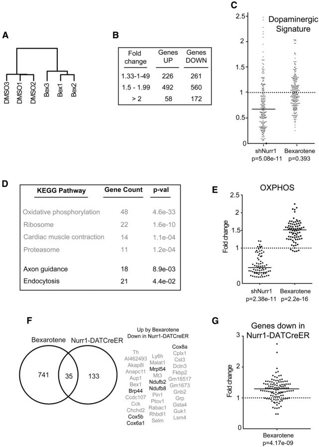
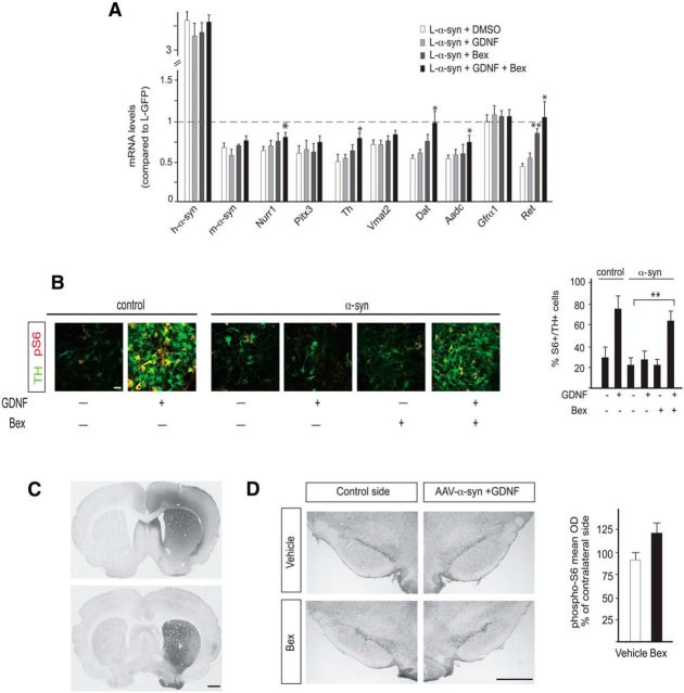
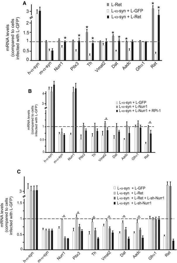
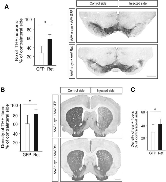
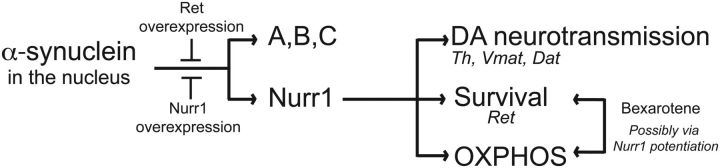
α-synuclein, a protein enriched in Lewy bodies and highly implicated in neurotoxicity in Parkinson's disease, is distributed both at nerve terminals and in the cell nucleus. Here we show that a nuclear derivative of α-synuclein induces more pronounced changes at the gene expression level in mouse primary dopamine (DA) neurons compared to a derivative that is excluded from the nucleus. Moreover, by RNA sequencing we analyzed the extent of genome-wide effects on gene expression resulting from expression of human α-synuclein in primary mouse DA neurons. The results implicated the transcription factor Nurr1 as a key dysregulated target of α-synuclein toxicity. Forced Nurr1 expression restored the expression of hundreds of dysregulated genes in primary DA neurons expressing α-synuclein, and therefore prompted us to test the possibility that Nurr1 can be pharmacologically targeted by bexarotene, a ligand for the retinoid X receptor that forms heterodimers with Nurr1. Although our data demonstrated that bexarotene was ineffective in neuroprotection in rats in vivo, the results revealed that bexarotene has the capacity to coregulate subsets of Nurr1 target genes including the receptor tyrosine kinase subunit Ret. Moreover, bexarotene was able to restore dysfunctional Ret-dependent neurotrophic signaling in α-synuclein-overexpressing mouse DA neurons. These data highlight the role of the Nurr1-Ret signaling pathway as a target of α-synuclein toxicity and suggest that retinoid X receptor ligands with appropriate pharmacological properties could have therapeutic potential in Parkinson's disease.

Significance statement: How α-synuclein, a protein enriched in Lewy bodies in Parkinson's disease, is causing neuropathology in dopamine neurons remains unclear. This study elucidated how α-synuclein is influencing gene expression and how Nurr1, a transcription factor known to protect dopamine neurons against α-synuclein toxicity, can counteract these effects. Moreover, given the protective role of Nurr1, this study also investigated how Nurr1 could be pharmacologically targeted via bexarotene, a ligand of Nurr1's heterodimerization partner retinoid X receptor (RXR). The results showed that RXR ligands could increase neurotrophic signaling, but provided a mixed picture of its potential in a Parkinson's disease rat model in vivo. However, this study clearly emphasized Nurr1's neuroprotective role and indicated that other RXR ligands could have therapeutic potential in Parkinson's disease.


Figures

Figure 1.
Figure 1.
Effects of WT, cytoplasmic (NES), and nuclear (NLS) α-synuclein on DA neuron marker expression. A–D, E13.5 mouse primary ventral midbrain cultures. A, qPCR analysis of α-synuclein, Nurr1, Pitx3, and Nurr1 target genes after infection with α-synuclein/GFP lentiviruses. All data are represented as mean + SEM of the fold change normalized against Rpl19 levels; n = 3. *p < 0.05. B, Immunostaining showing α-synuclein and TH protein expression in primary cultures from dissected E13.5 ventral midbrains at the indicated time points postinfection (p.i.) with α-synuclein or control lentiviruses. The arrowheads point to fragmented dendrites. The diagram on the right shows the number of TH+ neuronal soma per counted field (mean values + SEM; n = 3). ***p < 0.001. C, Expression of TH and α-synuclein analyzed by immunohistochemistry. Cultures were infected with control or α-synuclein lentiviruses. D, Western blot analysis of α-synuclein in cytoplasmic (left) and nuclear (right) cell extracts from E13.5 mouse primary ventral midbrain cultures infected for 5 d with the indicated lentiviruses. Tuj1 is used as a cytoplasmic marker and histone H3 as a nuclear marker. Scale bars: 50 μm.
Figure 2.
Figure 2.
Nurr1-mediated restoration of disrupted DA neuron gene expression after α-synuclein overexpression. A, qPCR analysis of α-synuclein, Nurr1, Pitx3, and selected DA neuron markers after infection with eGFP or the indicated Nurr1 lentiviruses. All data are represented as mean + SEM of the fold change normalized against Rpl19 levels; n = 3. B, Immunostaining showing α-synuclein and TH protein expression in primary cultures from dissected E13.5 ventral midbrains 5 d after infection with control, α-synuclein, or α-synuclein and Nurr1 lentiviruses. Scale bar, 50 μm. C, Western blot analysis of α-synuclein and actin in whole cell extracts from E13.5 mouse primary ventral midbrain cultures infected for 5 d with control, α-synuclein, and α-synuclein, and Nurr1 lentiviruses. D, Coimmunoprecipitation of overexpressed Nurr1 and α-synuclein (left) or Gal4-HA-RXR (positive control; right) in HEK293T cells. *p < 0.05; **p < 0.01.
Figure 3.
Figure 3.
RNA sequencing of Pitx3-eGFP primary midbrain DA cultures infected with α-synuclein or α-synuclein and Nurr1 lentiviruses. A, B, Volcano plots showing all genes (A) or DA signature genes (B) with RPKM values >1 after infection of E13.5 primary ventral midbrain cultures with α-synuclein or α-synuclein and Nurr1 lentiviruses. The negative log10 transformed p values (y-axis) test the null hypothesis of no difference in expression levels and are plotted against the average log2 fold changes in expression (x-axis). Genes in yellow are differentially expressed with an absolute log2 fold change >1. C, Tables showing the DA signature genes significantly upregulated/downregulated after α-synuclein overexpression.
Figure 4.
Figure 4.
RNA sequencing of FACS-sorted Pitx3-eGFP DA neurons treated with bexarotene or vehicle. A, Cluster dendrogram showing the similarity between the different RNASeq samples based on their Spearman correlation. B, Table showing the number of genes with an RPKM value >1 that are upregulated or downregulated by bexarotene (padj < 0.05). C, Scatter diagram showing the fold change of the RPKM values for the DA signature genes after infection with shNurr1 lentivirus or treatment with bexarotene. Bar shows the median value. D, Table showing the gene count and p values (after multiple hypothesis testing with the Benjamini–Hochberg method) for the significantly upregulated and downregulated KEGG (Kyoto Encyclopedia of Genes and Genomes) pathways in bexarotene-treated DA neurons. E, Scatter diagram showing the fold change of the RPKM values for oxidative phosphorylation genes after infection with shNurr1 lentivirus or treatment with bexarotene. Bars show median values. F, Left, Venn diagram showing the overlap between the genes significantly (padj < 0.05) upregulated by bexarotene treatment and the genes significantly (padj < 0.05) downregulated in DA neurons of Nurr1-DATCreER animals. Right, Table with the 35 overlapping genes with genes encoding proteins with a function in mitochondria being highlighted. G, Scatter diagram showing the fold change of the RPKM values by bexarotene for the genes significantly downregulated in Nurr1-DATCreER animals. Bar shows the median value.
Figure 5.
Figure 5.
Effect of bexarotene on Nurr1-dependent gene expression, DA neuron cell loss, and motor performance in the 6-OHDA and AAV-α-synuclein rat PD models. A, qPCR analysis of Nurr1, Pitx3, and Nurr1 target genes in the ventral midbrain of rats after 6-OHDA injection and/or daily treatment for 1 week with vehicle or two doses of bexarotene (1 and 3 mg/kg) by oral gavage. Levels of mRNAs in intact rats are set at 1. n = 4. *p < 0.05. B, TH immunostaining in the substantia nigra of rats receiving 6-OHDA plus vehicle or daily 3 mg/kg bexarotene by oral gavage and stereological counts of TH+ neurons in the substantia nigra. C, TH immunostaining in the striatum of rats receiving 6-OHDA plus vehicle or daily 3 mg/kg bexarotene by oral gavage and optical densitometry of TH+ striatal fibers. n = 8 per group. D, Motor performance in the amphetamine rotation (left) and the cylinder tests (right) in rats injected unilaterally with 6-OHDA and treated with vehicle or bexarotene (3 mg/kg) by oral gavage. n = 8 per group. E, Optical densitometry of TH+ striatal fibers and stereological counts of TH+ neurons in the substantia nigra of rats injected unilaterally with AAV-α-synuclein and treated with vehicle or bexarotene (10 mg/kg) by oral gavage. n = 7–8 per group. F, Motor performance in the cylinder test in rats injected unilaterally with AAV-α-synuclein and treated with vehicle or bexarotene (10 mg/kg) by oral gavage. n = 7–8 per group. Mean values + SEM are shown for all panels. *p < 0.05. Scale bars: 1 cm.
Figure 6.
Figure 6.
A, B, Potentiation of the neuroprotective effect of GDNF by bexarotene. Primary ventral midbrain cultures of E13.5 mouse embryos. A, qPCR analysis of Nurr1, Pitx3, and Nurr1 target genes after infection with α-synuclein lentivirus and treatment with DMSO, bexarotene (100 nm), GDNF (30 ng/ml), or GDNF together with bexarotene. Levels of mRNAs in cultures infected with eGFP lentivirus are set at 1. Mean values + SEM are shown; n = 3. B, Expression of TH and phospho-S6 analyzed by immunohistochemistry. Cultures were infected with control or α-synuclein lentivirus and treated with DMSO, bexarotene (100 nm), GDNF (30 ng/ml), or GDNF together with bexarotene. The diagram on the right shows the percentage of TH+ neurons expressing phospho-S6. Mean values + SEM are shown; n = 3. C, Immunohistochemistry showing the spread of GDNF after striatal injection of the recombinant peptide and the expression of human α-synuclein in the striatum resulting from nigral delivery of the AAV-α-synuclein vector. D, Immunohistochemistry showing the expression of phospho-S6 in rats receiving AAV-α-synuclein and GDNF plus daily bexarotene or vehicle. The diagram shows the optical density of phospho-S6 staining (mean density per cell) in the nigral TH+ neurons on the treated side, expressed as a percentage of the intensity measured on the noninjected side (n = 5 per group). Scale bars: B, 50 μm; C, D, 1 cm. *p < 0.05; **p < 0.01.
Figure 7.
Figure 7.
Ret restores DA marker expression after α-synuclein overexpression in vitro. A–C, qPCR analysis of α-synuclein, Nurr1, Pitx3, and Nurr1 target genes after infection of primary ventral midbrain cultures of E13.5 mouse embryos with the indicated lentiviruses, in the presence or absence of the Ret kinase inhibitor RPI-1 (B). Levels of mRNAs in cultures infected with eGFP lentivirus are set at 1. All data is represented as mean + SEM of the fold change normalized against Rpl19 levels; n = 3. *p < 0.05.
Figure 8.
Figure 8.
Ret protects nigral DA neurons against α-synuclein toxicity in vivo. A, Stereological counts of substantia nigra TH+ neurons (left) performed on TH-immunostained midbrain sections (right). B, Optical densitometry of the striatal TH+ innervation (left) performed on TH-immunostained striatal sections (right). C, Optical densitometry of the striatal rat α-synuclein+ innervation. Scale bars: 1 cm. *p < 0.05.
Figure 9.
Figure 9.
Protection from nuclear α-synuclein-induced toxicity by Nurr1/Ret overexpression and/or bexarotene treatment. Nuclear α-synuclein interferes with gene expression by targeting Nurr1 and other genes (A,B,C). Nurr1 affects the expression of genes involved in DA neurotransmission, in survival via the GDNF-Ret pathway and in OXPHOS. Nurr1 and Ret overexpression protect from the toxic effects of α-synuclein, whereas bexarotene restores Ret expression in α-synuclein overexpressing neurons and upregulates OXPHOS.

Similar articles

Cited by

References

    1. Anders S, Huber W. Differential expression analysis for sequence count data. Genome Biol. 2010;11:R106. doi: 10.1186/gb-2010-11-10-r106. - DOI - PMC - PubMed
    1. Anichtchik O, Calo L, Spillantini MG. Synaptic dysfunction in synucleinopathies. CNS Neurol Disord Drug Targets. 2013;12:1094–1100. - PubMed
    1. Barceló-Coblijn G, Golovko MY, Weinhofer I, Berger J, Murphy EJ. Brain neutral lipids mass is increased in alpha-synuclein gene-ablated mice. J Neurochem. 2007;101:132–141. - PubMed
    1. Bar-On P, Crews L, Koob AO, Mizuno H, Adame A, Spencer B, Masliah E. Statins reduce neuronal alpha-synuclein aggregation in in vitro models of Parkinson's disease. J Neurochem. 2008;105:1656–1667. doi: 10.1111/j.1471-4159.2008.05254.x. - DOI - PMC - PubMed
    1. Benjamini Y, Hochberg Y. Controlling the false discovery rate: a practical and powerful approach to multiple testing. J R Statist Soc B. 1995;57:289–300.

Publication types

MeSH terms