. 2015 Oct 21;35(42):14406-22.
doi: 10.1523/JNEUROSCI.1468-15.2015.

Reversible Deactivation of Motor Cortex Reveals Functional Connectivity with Posterior Parietal Cortex in the Prosimian Galago (Otolemur garnettii)

Affiliations

Reversible Deactivation of Motor Cortex Reveals Functional Connectivity with Posterior Parietal Cortex in the Prosimian Galago (Otolemur garnettii)

Dylan F Cooke et al. J Neurosci. .

Abstract

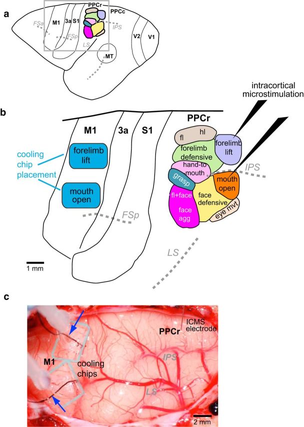
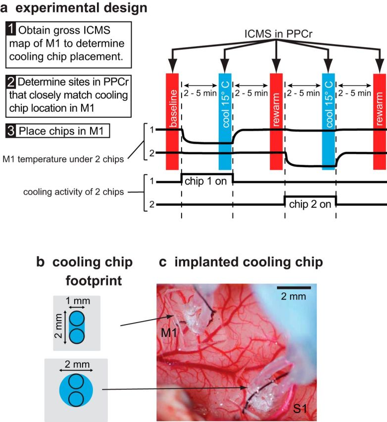
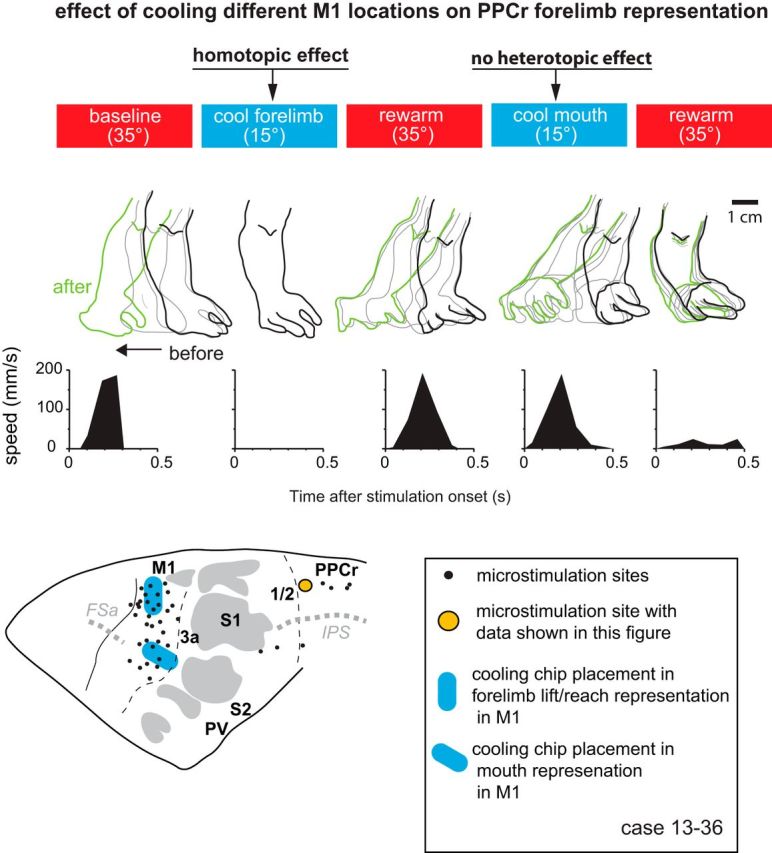
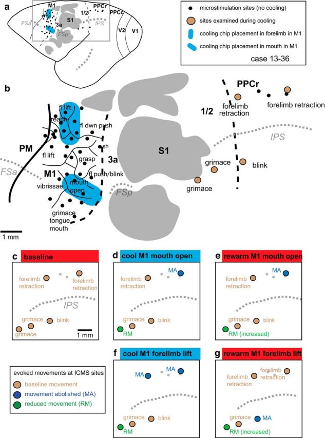
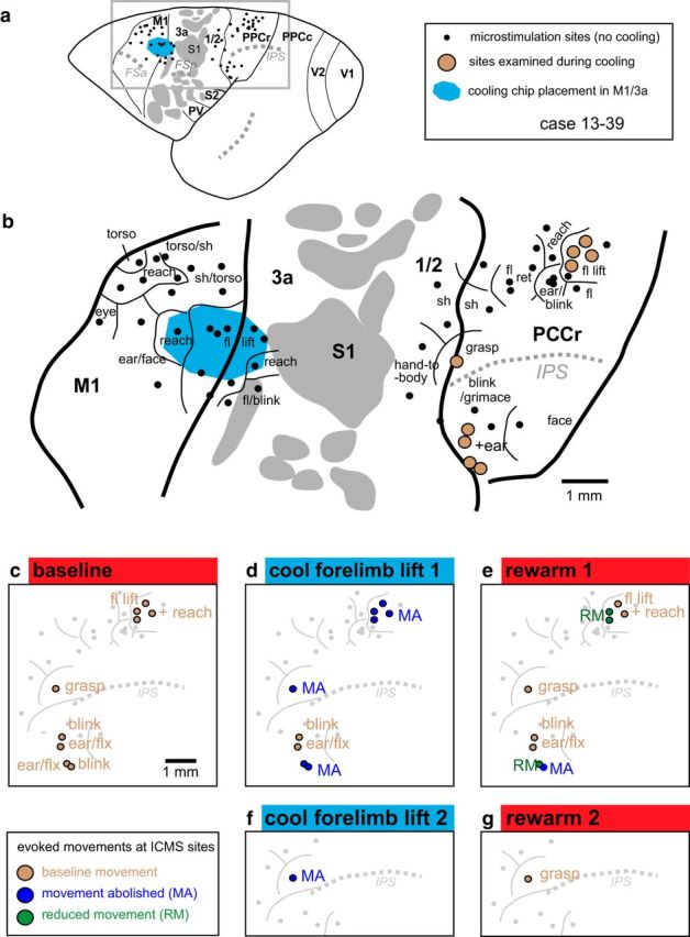
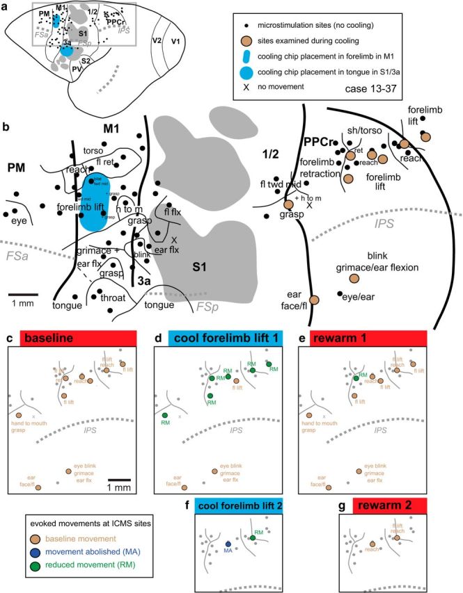
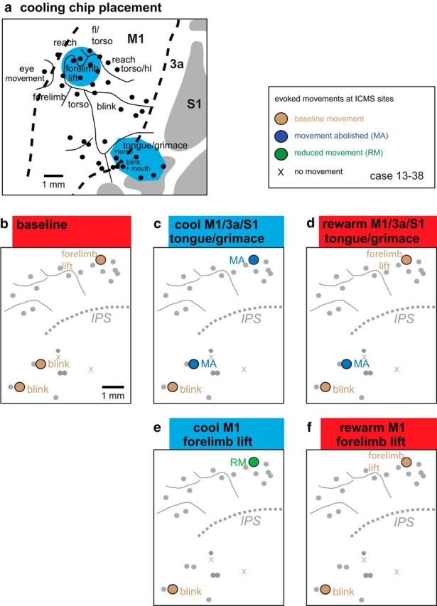
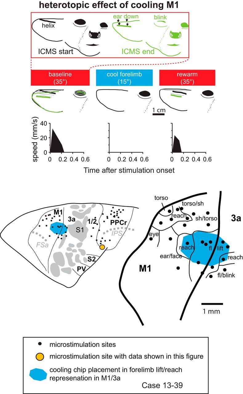
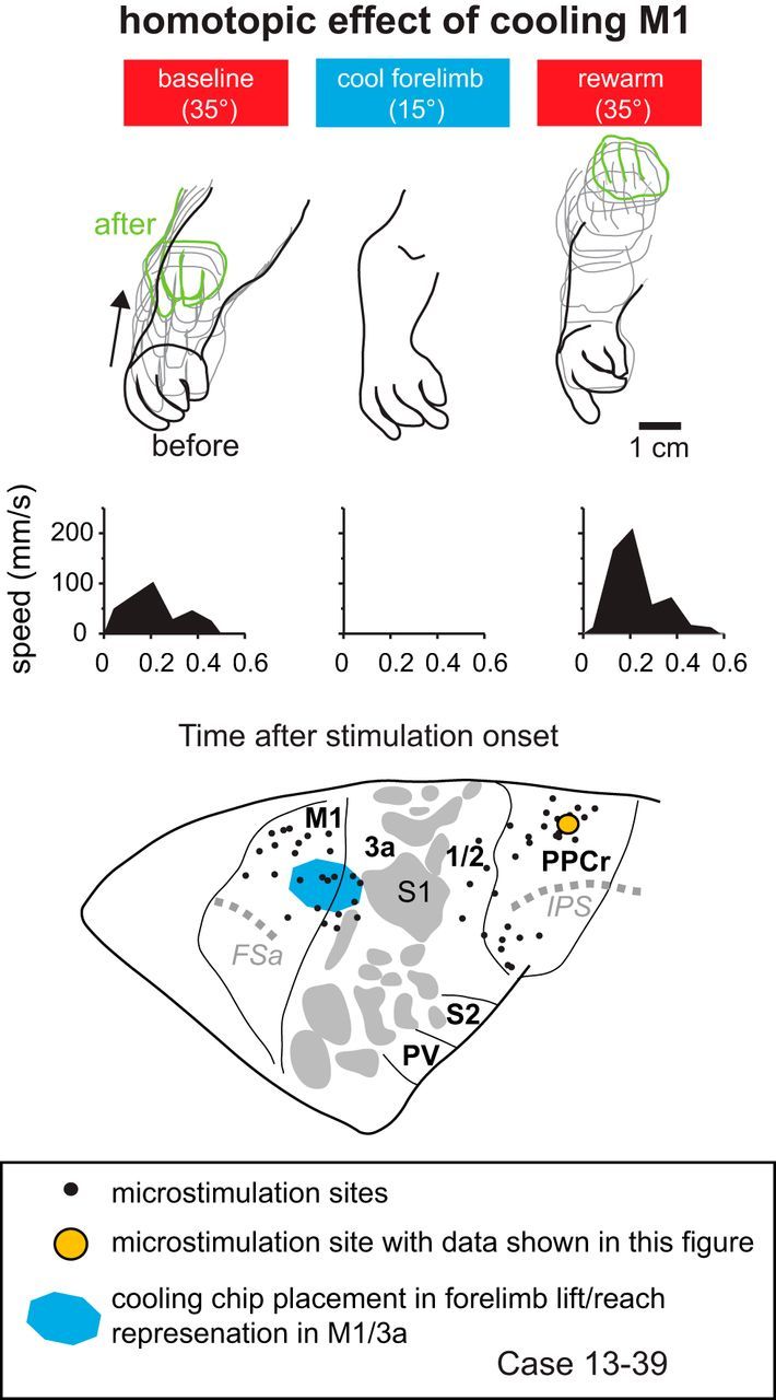
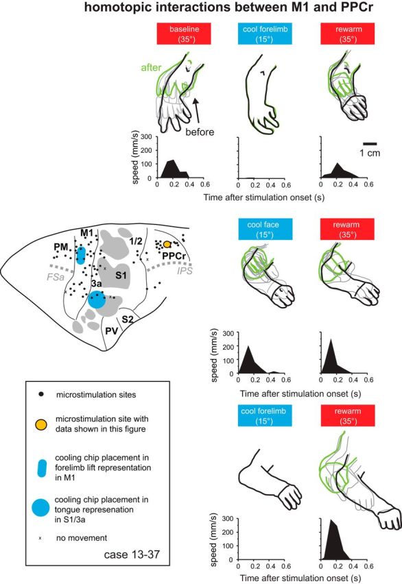
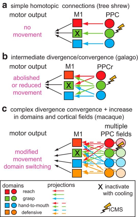
We examined the functional macrocircuitry of frontoparietal networks in the neocortex of prosimian primates (Otolemur garnettii) using a microfluidic thermal regulator to reversibly deactivate selected regions of motor cortex (M1). During deactivation of either forelimb or mouth/face movement domains within M1, we used long-train intracortical microstimulation techniques to evoke movements from the rostral division of posterior parietal cortex (PPCr). We found that deactivation of M1 movement domains in most instances abolished movements evoked in PPCr. The most common effect of deactivating M1 was to abolish evoked movements in a homotopic domain in PPCr. For example, deactivating M1 forelimb lift domains resulted in loss of evoked movement in forelimb domains in PPCr. However, at some sites, we also observed heterotopic effects; deactivating a specific domain in M1 (e.g., forelimb lift) resulted in loss of evoked movement in a different movement domain in PPCr (e.g., hand-to-mouth or eye-blink). At most sites examined in PPCr, rewarming M1 resulted in a reestablishment of the baseline movement at the same amplitude as that observed before cooling. However, at some sites, reactivation did not result in a return to baseline movement or to the full amplitude of the baseline movement. We discuss our findings in the context of frontoparietal circuits and how they may subserve a repertoire of ecologically relevant behaviors.

Significance statement: The posterior parietal cortex (PPC) of primates integrates sensory information used to guide movements. Different modules within PPC and motor cortex (M1) appear to control various motor behaviors (e.g., reaching, defense, and feeding). How these modules work together may vary across species and may explain differences in dexterity and even the capacity for tool use. We investigated the functional connectivity of these modules in galagos, a prosimian primate with relatively simple frontoparietal circuitry. By deactivating a reaching module in M1, we interfered with the function of similar PPC modules and occasionally unrelated PPC modules as well (e.g., eye blink). This circuitry in galagos, therefore, is more complex than in nonprimates, indicating that it has been altered with the expansion of primate PPC.

Keywords: cooling inactivation; lesion; muscimol; network; primate; reaching.


Figures

Figure 1.
Figure 1.
a, Dorsolateral view of the galago neocortex depicting major sulci (italicized abbreviations) and the positions of cortical areas relative to these sulci. The black box, enclosing M1, PPCr, and other fields examined here, indicated the region shown in b. b, In this hypothetical example, after exploration of both M1 and PPCr with long-train ICMS, cooling chips (blue rectangles) were implanted over the forelimb-lift and mouth-open domains in motor cortex. In PPCr, representations that corresponded to those over which the cooling chips were implanted were explored using long-train ICMS before, during, and after thermal deactivation in M1. Baseline movements evoked by ICMS in PPCr were compared with those evoked while the forelimb lift or the mouth representation in M1 were cooled to 10–15°C and to movements evoked after M1 temperature and function were restored. c, Digital image of the exposed neocortex with cooling chips implanted in M1 and a stimulation electrode inserted into PPCr from case 13–36. As the cooling chips are transparent, the outer margin of the PDMS bodies are outlined in gray. The smaller cooling footprint is opaque. Microthermocouples (blue arrows) are embedded in the cooling chips so that cortical temperature can be monitored continuously in both cooling chips throughout the experiment. This image was taken 12 h after implantation and after multiple cooling and warming epochs. Note that the cortex below the cooling chip has normal vasculature and no damage has been induced by this procedure. Maps of PPCr in a and b are modified from Stepniewska et al., 2005. See Table 1 for abbreviations.
Figure 2.
Figure 2.
Schematic of our experimental design (a), dimensions of cooling chip footprint (b), and implantation in M1 and S1 in a galago (c). a, Experiments started with gross ICMS maps of M1 (Step 1) and PPCr (Step 2). Based on these data, cooling chips were placed at one or two locations in M1 (Step 3). For each ICMS site, we tested stimulation during three epochs (vertical colored bars): baseline, cool (deactivation in M1 or S1), and rewarm (recovery). When two chips had been placed, there were two additional ICMS epochs tested: cooling of the second chip and rewarm. During each epoch, we studied movements evoked by long-train ICMS in PPCr. There was a 2–5 min interepoch interval such that ICMS data collection occurred after the cooled tissue had been stabilized for several minutes at a given temperature. Top black traces, Temperature of cortical surface under each cooling chip. Bottom black traces, Timing of cooling for each chip. Cooling devices have a small footprint (b) and are largely transparent when implanted on the cortex (c). The footprints illustrated in c are those of the cooling chips implanted in c over the forelimb lift representation in M1 and the tongue representation in S1 (case 13–37). In c, medial is to the top and rostral is to the right. Conventions are as in previous figure.
Figure 3.
Figure 3.
a, Digital image of a single section of cortex that was flattened, sectioned tangential to the cortical surface, and stained for CO in case 13–39. Area 3b stains heterogeneously, containing CO-dark and CO-light regions. Area 3a is lightly stained and M1 and area 1/2 stain moderately for CO. b, Reconstruction from the same case generated from the entire series of CO sections. It should be noted that individual sections reveal only some of the cortical field boundaries and an entire series combined with a series of additional stains such as myelin are used to reconstruct all cortical field boundaries. Thick black lines mark cortical field boundaries. Dotted gray lines are sulci. Myelin/CO dark islands in S1 are shaded gray. Rostral is to the right and medial is to the top. Conventions are as in previous figures.
Figure 4.
Figure 4.
Effect of cooling motor cortex on PPCr stimulation-evoked movements in case 13–36. Arm movements evoked by stimulating a site in PPCr (orange circle) were videotaped before, during, and after cooling deactivation of the forelimb lift/reach or tongue representations in M1. Stimulation epochs are represented by red (baseline warm and rewarm) and blue (cool) rectangles at the top. The outline of the forelimb has been traced for individual video frames during the evoked movement for each epoch. Black tracing is forelimb position before the movement, green is position after the movement, and gray is intermediate positions. Plots illustrate forelimb speed. Cooling the forelimb lift representation in M1 abolished evoked movements in the forelimb lift representation in PPCr, categorized here as a homotopic effect (affecting the same limb). Cooling the M1 tongue representation did not affect evoked movements in the forelimb representation in PPCr; that is, at this site, there was not a heterotopic effect from M1 face to PPCr forelimb.
Figure 5.
Figure 5.
Reconstruction of case 13–36. a, Location of IMCS sites in M1 and PPCr and the placement and size of cooling chips relative to these sites, major sulci, and cortical fields in a whole brain. b, Enlargement of boxed area in a with added ICMS maps obtained in M1 and PPCr before cooling regions in M1. In this case, a cooling chip was placed over forelimb representations in M1 (lift, reach, downward push). A second cooling chip was placed over the mouth-open representation in M1. Similar representations were identified in PPCr. cg, Effects in PPCr during cooling and rewarming of the forelimb and mouth representations in M1. c, Baseline evoked movements in PPCr. d, Cooling deactivation of the mouth representation in M1 affects sites in both the mouth (grimace) and the forelimb representation in PPCr. e, Rewarming the mouth representation in M1 resulted in a return of the evoked movement in the mouth representation (although reduced), whereas the evoked movement in the forelimb representation did not return to the baseline condition. Evoked movements were tested at these same sites when the forelimb lift representation in M1 was cooled (f) and then rewarmed (g). Cooling the forelimb lift representation in M1 abolished evoked movements in the forelimb lift representation in PPCr and reduced evoked movement at one site in the face representation of PPCr. Rewarming this representation in M1 returned movements to normal within the forelimb representation in PPCr. The evoked movement at the one affected site in the face representation increased in amplitude with rewarming, but did not return to baseline levels. Thick black lines mark cortical field boundaries (dashed black lines represent estimated boundaries). Thin black lines mark boundaries of movement representations. Dotted gray lines are sulci. Myelin/CO-dark islands in S1 are shaded gray. Conventions are as in previous figures. See Table 1 for abbreviations.
Figure 6.
Figure 6.
Reconstruction of case 13–39. a, Location of IMCS sites, cooling chip, major sulci, and cortical fields. b, Enlargement of boxed area in a. In this case, the cooling chip was placed over forelimb lift representations in M1, but also covered a small portion of area 3a. ce, Effects in PPCr during cooling and rewarming of the forelimb lift representation in M1. The effects of cooling the forelimb lift representation in M1 were most profound for similar movement representations (e.g., fl lift, reach) in PPCr, although sites that evoked a grasp movement were also affected (see red and blue sites in the medial portion of ce). Interestingly, cooling of the reach, fl lift representations in M1 also affected heterotopic representations in PPCr (e.g., eye blink and ear flexion), but this effect was only for some stimulation sites (see red and blue sites in the lateral portion of ce). At one site (grasp), we cooled and rewarmed twice (f, g). We found the same affect (extinction of evoked movement) on both the first and second cooling epochs (d, f), as well as a return of movement both times that cortex was returned to its normal temperature (e, g). See Table 1 for abbreviations. Conventions are as in previous figures.
Figure 7.
Figure 7.
Reconstruction of case 13–37. a, Location of IMCS sites, cooling chips, major sulci, and cortical fields. b, Enlargement of boxed area in a. In this case, the cooling chips were placed over forelimb lift representation in M1 and the tongue representation in S1/3a. Cooling the tongue representation had no effect on any of our tested sites in PPCr. ce, Effects in PPCr during cooling and rewarming of the forelimb lift representation in M1. Here, cooling in M1 affected all related PPCr sites (forelimb lift, reach, hand to mouth; d) except one and affected no unrelated sites (e.g., eye blink, ear flexion; d). In this case, at two sites, reduction or abolition of evoked movements were replicated in a second cooling and rewarming cycle (f, g). Many evoked movements in both M1 and PPCr were more complex than described in bg and included smaller movements of other body parts. However, for clarity in illustrating, these are only noted for sites under the cooling chip and for sites tested in PPCr. Conventions are as in previous figures.
Figure 8.
Figure 8.
Reconstructions of data from case 13–38. a, Enlargement of the area in which cooling chips were placed in the M1-forelimb-lift representation and the tongue/grimace representation in M1/3a/S1. The results of cooling and rewarming these representation on evoked movement in PPCr are illustrated in bf. Conventions are as in previous figures.
Figure 9.
Figure 9.
Effect of cooling motor cortex on PPCr stimulation-evoked movements in case 13–39. At top, the face is shown before and just after ICMS during the baseline epoch. Downward ear movement and unilateral eye blink evoked by stimulating a site in PPCr (orange circle in cortical map at bottom) were videotaped before, during, and after cooling deactivation of the forelimb lift representation in M1/3a (second row of tracings). Plots show speed of the helix of the pinna (traced with thick lines), the most easily tracked feature of the ear from this video angle. This figure demonstrates heterotopic interactions between M1 and PPCr in which an unrelated movement evoked in PPCr is abolished. When cortex was returned to normal temperature, the ear movement returned, although the blink did not. Conventions are as in previous figures.
Figure 10.
Figure 10.
Effect of cooling motor cortex on PPCr stimulation-evoked movements in case 13–39. Arm movements evoked by stimulating a site in PPCr (orange circle) were videotaped before, during, and after cooling deactivation of the forelimb lift representation in M1/3a. This figure demonstrates homotopic interactions between M1 and PPCr in which a similar movement evoked in PPCr is abolished. When cortex was returned to normal temperature, the movement returned, the amplitude of the movement was greater, and the end position of the movement was higher than baseline. Conventions are as in previous figures.
Figure 11.
Figure 11.
Effect of cooling motor cortex on PPCr stimulation-evoked movements in case 13–37. Arm movements evoked by stimulating a site in PPCr (orange circle) were videotaped before, during, and after cooling deactivation of the forelimb lift representation in M1 and the tongue representation in 3a/S1. The top row shows results when cooling and rewarming the forelimb lift representation in M1, the middle row shows the results when cooling and rewarming the tongue representation in area 3a/S1 (“cool face”), and the bottom row shows the results M1 was cooled and rewarmed a second time. For both M1-forelimb cooling cycles (first and third rows), stimulation movement was nearly or completely abolished during cooling of the M1 forelimb lift representation and recovered after rewarming of M1. Cooling the tongue representation in areas 3a/S1 had no effect on evoked movements at this site. The brain at the left shows the position of cooling chips and the ICMS in PPC in this case. Conventions are as in previous figures.
Figure 12.
Figure 12.
Schematic illustrating simple (a), intermediate (b), and complex (c) motor-posterior parietal networks. Nondivergent, homotopic projections from PPC to M1 are hypothesized for tree shrews (as in a) and divergent projections from movement domains in different areas in PPC to M1 are hypothesized for the macaque monkey (c). For example, when a given domain in M1 is deactivated via cooling (e.g., X in grasp domain in M1), in a simple nondivergent network (a) stimulation of a similar domain in PPC would result in no movements. A very complex network consisting of divergent and convergent connectivity from multiple domains and multiple cortical fields (c) would have a different motor output, perhaps including domain switching or modified movements. We hypothesize that galagos have networks of intermediate complexity (b).

Similar articles

Cited by

References

    1. Aihara H, Okada Y, Tamaki N. The effects of cooling and rewarming on the neuronal activity of pyramidal neurons in guinea pig hippocampal slices. Brain Res. 2001;893:36–45. doi: 10.1016/S0006-8993(00)03285-6. - DOI - PubMed
    1. Andersen RA, Andersen KN, Hwang EJ, Hauschild M. Optic ataxia: from Balint's syndrome to the parietal reach region. Neuron. 2014;81:967–983. doi: 10.1016/j.neuron.2014.02.025. - DOI - PMC - PubMed
    1. Asanuma H, Rosén I. Topographical organization of cortical efferent zones projecting to distal forelimb muscles in the monkey. Exp Brain Res. 1972;14:243–256. doi: 10.1007/BF00816161. - DOI - PubMed
    1. Bakola S, Passarelli L, Gamberini M, Fattori P, Galletti C. Cortical connectivity suggests a role in limb coordination for macaque area PE of the superior parietal cortex. J Neurosci. 2013;33:6648–6658. doi: 10.1523/JNEUROSCI.4685-12.2013. - DOI - PMC - PubMed
    1. Baldwin MK, Cooke DF, Gordon A, Krubitzer L. Revealing functional organization of frontoparietal networks in tree shrews (Tupaia belangeri) using reversible inactivation. Soc Neurosci Abstr. 2014;40:446.02.

Publication types