. 2015 Dec 25;290(52):30888-900.
doi: 10.1074/jbc.M115.688630. Epub 2015 Oct 21.

A Second β-Hexosaminidase Encoded in the Streptococcus pneumoniae Genome Provides an Expanded Biochemical Ability to Degrade Host Glycans

Affiliations

A Second β-Hexosaminidase Encoded in the Streptococcus pneumoniae Genome Provides an Expanded Biochemical Ability to Degrade Host Glycans

Melissa Robb et al. J Biol Chem. .

Abstract

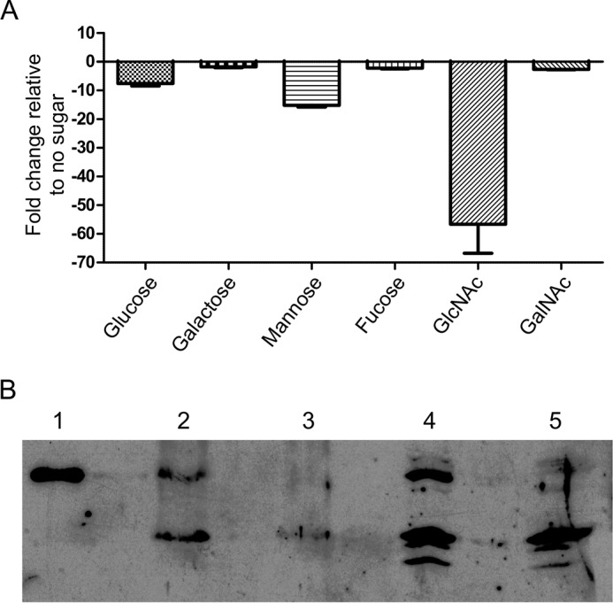
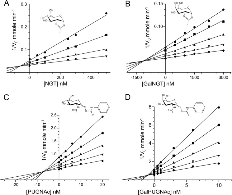
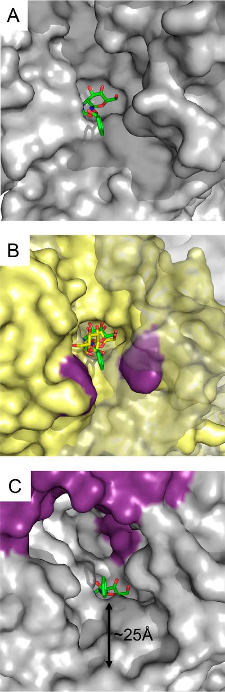
An important facet of the interaction between the pathogen Streptococcus pneumoniae (pneumococcus) and its human host is the ability of this bacterium to process host glycans. To achieve cleavage of the glycosidic bonds in host glycans, S. pneumoniae deploys a wide array of glycoside hydrolases. Here, we identify and characterize a new family 20 glycoside hydrolase, GH20C, from S. pneumoniae. Recombinant GH20C possessed the ability to hydrolyze the β-linkages joining either N-acetylglucosamine or N-acetylgalactosamine to a wide variety of aglycon residues, thus revealing this enzyme to be a generalist N-acetylhexosaminidase in vitro. X-ray crystal structures were determined for GH20C in a ligand-free form, in complex with the N-acetylglucosamine and N-acetylgalactosamine products of catalysis and in complex with both gluco- and galacto-configured inhibitors O-(2-acetamido-2-deoxy-D-glucopyranosylidene)amino N-phenyl carbamate (PUGNAc), O-(2-acetamido-2-deoxy-D-galactopyranosylidene)amino N-phenyl carbamate (GalPUGNAc), N-acetyl-D-glucosamine-thiazoline (NGT), and N-acetyl-D-galactosamine-thiazoline (GalNGT) at resolutions from 1.84 to 2.7 Å. These structures showed N-acetylglucosamine and N-acetylgalactosamine to be recognized via identical sets of molecular interactions. Although the same sets of interaction were maintained with the gluco- and galacto-configured inhibitors, the inhibition constants suggested preferred recognition of the axial O4 when an aglycon moiety was present (Ki for PUGNAc > GalPUGNAc) but preferred recognition of an equatorial O4 when the aglycon was absent (Ki for GalNGT > NGT). Overall, this study reveals GH20C to be another tool that is unique in the arsenal of S. pneumoniae and that it may implement the effort of the bacterium to utilize and/or destroy the wide array of host glycans that it may encounter.

Keywords: Streptococcus; enzyme inhibitor; glycobiology; glycoside hydrolase; x-ray crystallography.


Figures

FIGURE 1.
FIGURE 1.
GH20C expression. A, fold changes of GH20C transcript were measured by quantitative RT-PCR after exposure of TIGR4 to different carbohydrates. The fold change relative to expression levels when the bacterium was not exposed to sugar was calculated, and standard deviations of three replicates are indicated on each bar. B, production of GH20C protein by S. pneumoniae was detected by Western blotting using a polyclonal antibody raised against recombinant GH20C. Wild-type and Δgh20C mutant of S. pneumoniae TIGR4 were grown for 6 h at 37 °C in AGCH media and harvested by centrifugation. The cells were then lysed by sonication, and the culture supernatant and lysed cellular fractions were analyzed by Western blotting. Lane 1, recombinant GH20C; lane 2, TIGR4 wild-type supernatant; lane 3, Δgh20C mutant supernatant; lane 4, TIGR4 wild-type lysate; lane 5, Δgh20C mutant lysate.
FIGURE 2.
FIGURE 2.
X-ray crystal structure of GH20C. A, GH20C monomer with the loops represented in gray and the domains represented in green schematic for the N-terminal domain, yellow for the central (β/α)8 domain, and purple for the C-terminal domain. B, GH20C dimer with one monomer colored as in A and the other monomer represented as a slate blue surface. C, overlay of the GH20C active site (yellow stick representation for side chains and transparent gray schematic representation of the backbone) with GcnA (cyan stick representation for side chains) in complex with NGT (salmon stick representation). The numbering refers to that of the GH20C polypeptide.
FIGURE 3.
FIGURE 3.
Structure of GH20C in complex with reaction products. A, maximum likelihood σA-weighted Fo − Fc electron density maps contoured at 2.5σ for GlcNAc (left; represented in orange sticks) and GalNAc (right; represented in green sticks). B, specific interactions between the GH20C active site (side chains represented as yellow sticks with the catalytic residues as orange sticks) in its engaged form and GlcNAc (in magenta stick representation). Waters involved in the hydrogen bonding network are shown as red spheres. The loop carrying the catalytic residues is represented as orange schematic. C, overlay of two GH20C monomers from the GlcNAc complex structure. The engaged form is shown in yellow for the protein and with GlcNAc as magenta sticks. The form of the protein with the catalytic loop retracted is shown in green with the GlcNAc shown as cyan sticks. Relative active site residues are shown as sticks. D, overlay of GH20C in complex with GlcNAc (orange sticks with black dashed lines for hydrogen bonds) and GalNAc (green sticks with green dashed lines for hydrogen bonds).
FIGURE 4.
FIGURE 4.
Inhibition of GH20C. Dixon plot analyses used to determine the inhibition constant of NGT (A), GalNGT (B), PUGNAc (C), and GalPUGNAc (D). Insets show the chemical structures of the inhibitors. The concentration of enzyme used were 100 nm (circle), 150 nm (square), 200 nm (triangle), 350 nm (inverted triangle), and 500 nm (diamond).
FIGURE 5.
FIGURE 5.
Structures of GH20C in complex with inhibitors. Maximum likelihood σA-weighted Fo − Fc electron density maps for NGT (A) and GalNGT (B) contoured at 2.5σ. C, overlay of the active site of GH20C bound to NGT (yellow sticks) and GalNGT (blue sticks). Hydrogen bonds are represented by green dashed lines for the NGT complex and dashed black lines for the GalNGT complex. D, maximum likelihood σA-weighted Fo − Fc electron density maps for GalPUGNAc contoured at 2.5σ. E, overlay of the active site of GH20C bound to Gal-PUGNAc (salmon sticks) and PUGNAc (cyan sticks) found in the engaged conformations of the enzyme. Hydrogen bonds are represented by dashed black lines for the PUGNAc complex structure and dashed green lines for the Gal-PUGNAc complex structure. F and G, maximum likelihood σA-weighted Fo − Fc electron density maps for PUGNAc contoured at 2.5σ showing PUGNAc when bound to the three retracted conformation GH20C molecules in the asymmetric unit (F) and when bound the single “engaged” conformation GH20C molecules in the asymmetric unit (G).
FIGURE 6.
FIGURE 6.
Surface contours of the GH20C active site. A, solvent-accessible surface of the active site of the GH20C monomer, with GalPUGNAc shown as green sticks, shows the open nature of the active site. B, overlay of GH20C (gray transparent surface with GalPUGNAc show in green sticks) with the GH20A module of StrH. The surface of GH20A is shown in yellow with critical aromatic amino acids flanking the entrance to the active site shown in purple. The β-d-GlcNAc-(1→2)-β-d-Man disaccharide bound to GH20A is shown as yellow sticks. C, solvent-accessible surface of the active site of the GH20C dimer, with GalPUGNAc shown as green sticks, shows the tunnel of the active site, which is ∼25 Å from the entrance to the subsite accommodating the glycon (−1 subsite).

References

    1. Bøhle L. A., Mathiesen G., Vaaje-Kolstad G., and Eijsink V. G. (2011) An endo-β-N-acetylglucosaminidase from Enterococcus faecalis V583 responsible for the hydrolysis of high-mannose and hybrid-type N-linked glycans. FEMS Microbiol. Lett. 325, 123–129 - PubMed
    1. Jiang Y.-L., Yu W.-L., Zhang J.-W., Frolet C., Di Guilmi A.-M., Zhou C.-Z., Vernet T., and Chen Y. (2011) Structural basis for the substrate specificity of a novel β-N-acetylhexosaminidase StrH protein from Streptococcus pneumoniae R6. J. Biol. Chem. 286, 43004–43012 - PMC - PubMed
    1. Prag G., Papanikolau Y., Tavlas G., Vorgias C. E., Petratos K., and Oppenheim A. B. (2000) Structures of chitobiase mutants complexed with the substrate di-N-acetyl-d-glucosamine: the catalytic role of the conserved acidic pair, aspartate 539 and glutamate 540. J. Mol. Biol. 300, 611–617 - PubMed
    1. Sumida T., Stubbs K. A., Ito M., and Yokoyama S. (2012) Gaining insight into the inhibition of glycoside hydrolase family 20 exo-β-N-acetylhexosaminidases using a structural approach. Org. Biomol. Chem. 10, 2607–2612 - PubMed
    1. Williams S. J., Mark B. L., Vocadlo D. J., James M. N., and Withers S. G. (2002) Aspartate 313 in the Streptomyces plicatus hexosaminidase plays a critical role in substrate-assisted catalysis by orienting the 2-acetamido group and stabilizing the transition state. J. Biol. Chem. 277, 40055–40065 - PubMed

Publication types

MeSH terms

Associated data