. 2016 Jan;26(1):402-413.
doi: 10.1093/cercor/bhv203. Epub 2015 Oct 21.

Maturation of Sensori-Motor Functional Responses in the Preterm Brain

Affiliations

Maturation of Sensori-Motor Functional Responses in the Preterm Brain

Alessandro G Allievi et al. Cereb Cortex. 2016 Jan.

Abstract

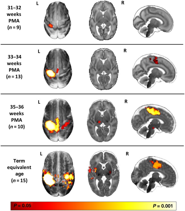
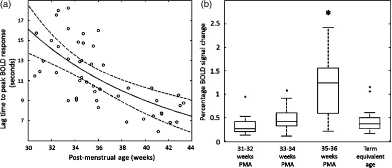
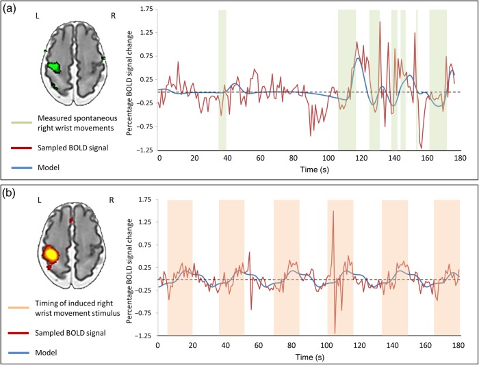
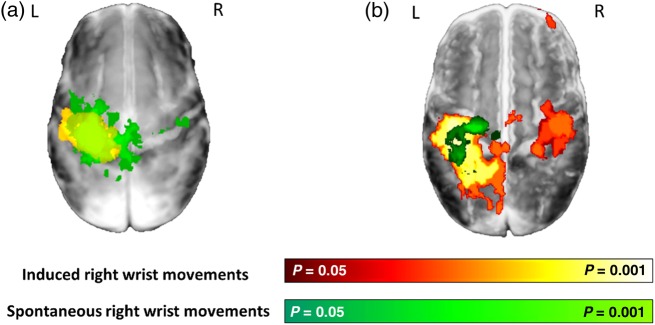
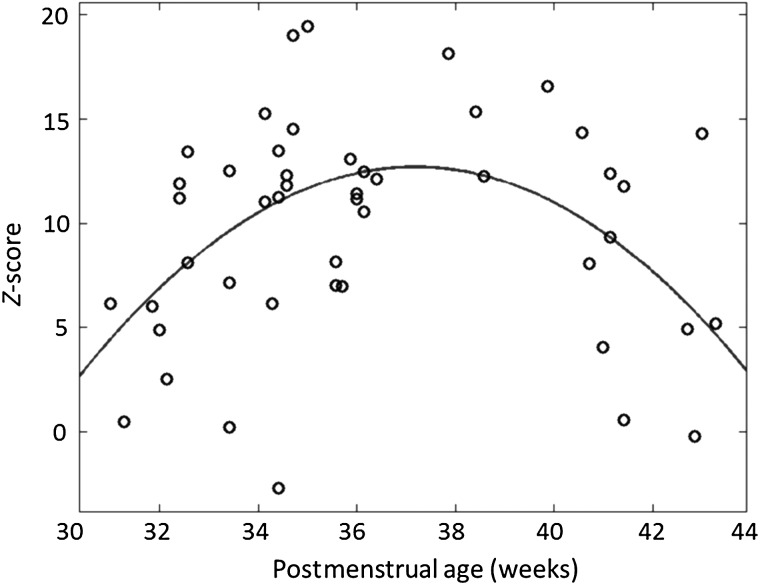
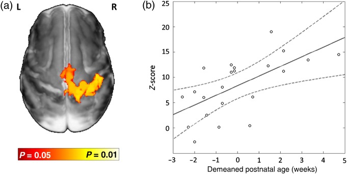
Preterm birth engenders an increased risk of conditions like cerebral palsy and therefore this time may be crucial for the brain's developing sensori-motor system. However, little is known about how cortical sensori-motor function matures at this time, whether development is influenced by experience, and about its role in spontaneous motor behavior. We aimed to systematically characterize spatial and temporal maturation of sensori-motor functional brain activity across this period using functional MRI and a custom-made robotic stimulation device. We studied 57 infants aged from 30 + 2 to 43 + 2 weeks postmenstrual age. Following both induced and spontaneous right wrist movements, we saw consistent positive blood oxygen level-dependent functional responses in the contralateral (left) primary somatosensory and motor cortices. In addition, we saw a maturational trend toward faster, higher amplitude, and more spatially dispersed functional responses; and increasing integration of the ipsilateral hemisphere and sensori-motor associative areas. We also found that interhemispheric functional connectivity was significantly related to ex-utero exposure, suggesting the influence of experience-dependent mechanisms. At term equivalent age, we saw a decrease in both response amplitude and interhemispheric functional connectivity, and an increase in spatial specificity, culminating in the establishment of a sensori-motor functional response similar to that seen in adults.

Keywords: development; neonate; sensori-motor; task fMRI.


Figures

Figure 1.
Figure 1.
Evolution of sensori-motor functional responses induced by passive movement of the right wrist during. Following right wrist movement, localized functional activity was identified in all infants in the contralateral (left) primary somatosensory cortex. Functional responses can be seen to progress from a contralateral only pattern in the youngest infants at 31–32 weeks postmenstrual age (PMA) (top row, n = 9); to include the midline supplementary motor area (SMA) at 33–34 weeks (second row, n = 13); and the ipsilateral peri-rolandic cortex and thalamus at 35–36 weeks (third row, n = 10). At term equivalent age (37–44 weeks; fourth row, n = 15), a mature adult-like activation pattern is seen in the bilateral peri-rolandic regions, basal ganglia, SMA, and contralateral opercular cortex/secondary somatosensory cortex. The images show the results of one-sample t-test performed using permutation testing and corrected for family-wise error (FWE) overlaid on an age-specific 4D brain T2-weighted brain atlas (Serag et al. 2012).
Figure 2.
Figure 2.
Maturational changes in the temporal characteristics and amplitude of the identified functional brain activity (a). The lag time to the positive peak of the identified response decreases systematically with increasing PMA (data shown with robust regression line with prediction error bands). (b) The amplitude of BOLD signal change within the identified clusters of activity are significantly higher at the conclusion of the third trimester (35–36 weeks PMA) in comparison to younger infants and those at term equivalent age (line represents data median; box represents data lower and upper quartile; *P < 0.05 Mann–Whitney U-test with Holm–Bonferroni correction).
Figure 3.
Figure 3.
Spontaneous limb movements in the preterm brain are associated with functional brain activity in the peri-rolandic cortex. (a) Using a precise fiber-optic position sensor, spontaneous “active” right wrist movements during image acquisition were recorded (green blocks) and convolved with an age-specific hemodynamic response function (HRF) to model (blue trace) the acquired BOLD signal (red trace). In this example, preterm infant (32 + 3 weeks PMA), spontaneous movements of the right wrist were significantly correlated with the mean BOLD signal in the left peri-rolandic cortex (green cluster). (b) This same approach can also be used to model brain responses induced by “passive” right wrist movement (orange blocks), with the measured BOLD signal closely fitting the design model. In the same infant, this allows the identification of a localized cluster of functional activity (red–yellow) in the left peri-rolandic region.
Figure 4.
Figure 4.
Maturation of functional brain activity associated with spontaneous motor behavior in infants during the preterm period and at term equivalent age. (a) Spontaneous movements of the right wrist in preterm infants (median age 33 + 3 weeks PMA, n = 10) were associated with a large cluster of activity (green) in the bilateral peri-rolandic regions which is nearly identical to that seen in the same infants following induced movement of the right wrist (red–yellow). (b) At term equivalent age (median age 41 weeks PMA, n = 8), the functional activity associated with spontaneous movements (green) is more focal and anterior to that seen following somatosensory stimulation (red–yellow).
Figure 5.
Figure 5.
Interhemispheric functional connectivity initially increases rapidly during the preterm period but then appears to decrease at term equivalent age. Partial correlation between the BOLD contrast time-series to somatosensory stimulation in the left and right peri-rolandic regions increases rapidly during the preterm period, and reaches a maximum value at 36 weeks PMA. It then falls at term equivalent age, leading to an inverted U-shaped distribution. This decrease in functional connectivity may result from increasingly specific functional connectivity. (Data represent z-transformed Pearson's partial correlation coefficient).
Figure 6.
Figure 6.
Interhemispheric functional connectivity during the preterm period is influenced by ex utero exposure. (a) Up to 35 weeks PMA (n = 22), functional activity following right wrist movement in the ipsilateral (right) peri-rolandic cortex is linearly related to the amount of ex utero exposure as measured by postnatal age. Image shows the results of a linear model fitting based on postnatal age at scan (and controlling for PMA) overlaid on an age-specific template. (b) In the same infants, functional connectivity between the BOLD signal time-series in the left and right peri-rolandic regions during the task also shows a linear relationship with the demeaned postnatal age.

References

    1. Aeby A, Liu Y, De Tiege X, Denolin V, David P, Baleriaux D, Kavec M, Metens T, Van Bogaert P. 2009. Maturation of thalamic radiations between 34 and 41 weeks’ gestation: a combined voxel-based study and probabilistic tractography with diffusion tensor imaging. AJNR Am J Neuroradiol. 30:1780–1786. - PMC - PubMed
    1. Allievi AG, Arichi T, Gordon AL, Burdet E. 2014. Technology-aided assessment of sensorimotor function in early infancy. Front Neurol. 5:197. - PMC - PubMed
    1. Allievi AG, Melendez-Calderon A, Arichi T, Edwards AD, Burdet E. 2013. An fMRI compatible wrist robotic interface to study brain development in neonates. Ann Biomed Eng. 41:1181–1192. - PubMed
    1. Als H, Duffy FH, McAnulty GB, Rivkin MJ, Vajapeyam S, Mulkern RV, Warfield SK, Huppi PS, Butler SC, Conneman N et al. . 2004. Early experience alters brain function and structure. Pediatrics. 113:846–857. - PubMed
    1. Anderson AW, Marois R, Colson ER, Peterson BS, Duncan CC, Ehrenkranz RA, Schneider KC, Gore JC, Ment LR. 2001. Neonatal auditory activation detected by functional magnetic resonance imaging. Magn Reson Imaging. 19:1–5. - PubMed

Publication types