. 2015 Oct 21;90(1):400-11.
doi: 10.1128/JVI.02082-15. Print 2016 Jan 1.

A Unique Multibasic Proteolytic Cleavage Site and Three Mutations in the HA2 Domain Confer High Virulence of H7N1 Avian Influenza Virus in Chickens

Affiliations

A Unique Multibasic Proteolytic Cleavage Site and Three Mutations in the HA2 Domain Confer High Virulence of H7N1 Avian Influenza Virus in Chickens

El-Sayed M Abdelwhab et al. J Virol. .

Abstract

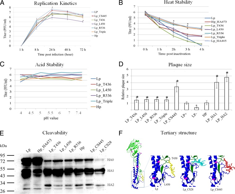
In 1999, after circulation for a few months in poultry in Italy, low-pathogenic (LP) avian influenza (AI) H7N1 virus mutated into a highly pathogenic (HP) form by acquisition of a unique multibasic cleavage site (mCS), PEIPKGSRVRR*GLF (asterisk indicates the cleavage site), in the hemagglutinin (HA) and additional alterations with hitherto unknown biological function. To elucidate these virulence-determining alterations, recombinant H7N1 viruses carrying specific mutations in the HA of LPAI A/chicken/Italy/473/1999 virus (Lp) and HPAI A/chicken/Italy/445/1999 virus (Hp) were generated. Hp with a monobasic CS or carrying the HA of Lp induced only mild or no disease in chickens, thus resembling Lp. Conversely, Lp with the HA of Hp was as virulent and transmissible as Hp. While Lp with a multibasic cleavage site (Lp_CS445) was less virulent than Hp, full virulence was exhibited when HA2 was replaced by that of Hp. In HA2, three amino acid differences consistently detected between LP and HP H7N1 viruses were successively introduced into Lp_CS445. Q450L in the HA2 stem domain increased virulence and transmission but was detrimental to replication in cell culture, probably due to low-pH activation of HA. A436T and/or K536R restored viral replication in vitro and in vivo. Viruses possessing A436T and K536R were observed early in the HPAI outbreak but were later superseded by viruses carrying all three mutations. Together, besides the mCS, stepwise mutations in HA2 increased the fitness of the Italian H7N1 virus in vivo. The shift toward higher virulence in the field was most likely gradual with rapid optimization.

Importance: In 1999, after 9 months of circulation of low-pathogenic (LP) avian influenza virus (AIV), a devastating highly pathogenic (HP) H7N1 AIV emerged in poultry, marking the largest epidemic of AIV reported in a Western country. The HPAIV possessed a unique multibasic cleavage site (mCS) complying with the minimum motif for HPAIV. The main finding in this report is the identification of three mutations in the HA2 domain that are required for replication and stability, as well as for virulence, transmission, and tropism of H7N1 in chickens. In addition to the mCS, Q450L was required for full virulence and transmissibility of the virus. Nonetheless, it was detrimental to virus replication and required A436T and/or K536R to restore replication, systemic spread, and stability. These results are important for better understanding of the evolution of highly pathogenic avian influenza viruses from low-pathogenic precursors.


Figures

FIG 1
FIG 1
Diagrams of the recombinant viruses generated in this study. Gene segments or mutations of the LP H7N1 virus are illustrated in blue, whereas those from the HP H7N1 virus are in red. The triangles in the HA indicate the cleavage site motif: blue (mCS motif PEIPKGR*G from Lp), red (mCS PEIPKGSRVRR*G from Hp), or yellow (mCS PEIPKRRRR*G from HP H7N7).
FIG 2
FIG 2
Clinical findings in chickens inoculated with H7N1 carrying different cleavage site motifs or HA proteins. Shown is clinical scoring after oculonasal inoculation of 4- to 6-week-old specific-pathogen-free White Leghorn chickens with 104.5 PFU/ml of the indicated viruses. Chickens without clinical signs were scored 0 (white boxes). A score of 1 (light gray boxes) was applied to chickens with one of the following clinical signs: depression, ruffled feathers, diarrhea, sneezing, coughing, conjunctivitis, discharges, or cyanosis of the comb, wattle, or shanks. These chickens were categorized as ill. Severely ill chickens showed two or more clinical signs and were scored 2 (dark gray boxes), whereas dead chickens were scored 3 (black boxes). The PI was calculated as the mean sum of the daily arithmetic mean values divided by 10, the number of observation days. “P” stands for pathology; the birds were killed and taken for necropsy.
FIG 3
FIG 3
Clinical findings in chickens inoculated with H7N1 carrying multibasic cleavage sites and specific mutations in the HA2 domain. Shown is clinical scoring after oculonasal inoculation of 4- to 6-week-old specific-pathogen-free White Leghorn chickens with 104.5 PFU/ml. Chickens without clinical illness were scored 0 (white boxes). A score of 1 (light gray boxes) was applied to chickens with one of the following clinical signs: depression, ruffled feathers, diarrhea, sneezing, coughing, conjunctivitis, discharges, or cyanosis of the comb, wattle, or shanks. These chickens were categorized as ill. Severely ill chickens showed two or more clinical signs and were scored 2 (dark gray boxes), whereas dead chickens were scored 3 (black boxes). The PI was calculated as the mean sum of the daily arithmetic mean values divided by 10, the number of observation days. “P” stands for pathology; the birds were killed and taken for necropsy.
FIG 4
FIG 4
Virus shedding and tissue tropism 4 days postinoculation of SPF chickens. (A) Mean amounts of virus excreted at day 4 postinoculation from the oral and cloacal swabs as estimated by plaque assay on MDCK cells. (B) Detection of influenza virus NP by immunohistochemistry in lungs, heart, and brain (bright red staining); data on the full tropism of all viruses are available upon request.
FIG 5
FIG 5
In vitro characterization of selected viruses reverse engineered in this study. (A) Replication kinetics as estimated by plaque test at 1, 8, 24, 48, and 72 h postinoculation of CEK cells at an MOI of 0.001 PFU/cell. (B) Heat stability at 50°C for 1, 2, 3, and 4 h. (C) Stability after incubation of viruses at pH 4, 4.5, 5, 5.5, 6, 7, or 7.4. (D) Sizes of plaques at MDCKII cells. (E) Cleavability of the HA of recombinant viruses 6 h after inoculation of CEK cells at an MOI of 1 in the absence of trypsin. (F) Predicted tertiary structure of the HA protein showing mutations in the HA of Lp versus Hp (pink) and variable cleavage site motifs (yellow in Lp, cyan in Lp_CS28, and red in Hp). The error bars represent the standard deviations of different replicates. The asterisks indicate P values of < 0.05.

Similar articles

Cited by

References

    1. Cheung TK, Poon LL. 2007. Biology of influenza A virus. Ann N Y Acad Sci 1102:1–25. doi:10.1196/annals.1408.001. - DOI - PubMed
    1. Brown EG. 2000. Influenza virus genetics. Biomed Pharmacother 54:196–209. doi:10.1016/S0753-3322(00)89026-5. - DOI - PubMed
    1. Tong S, Zhu X, Li Y, Shi M, Zhang J, Bourgeois M, Yang H, Chen X, Recuenco S, Gomez J, Chen LM, Johnson A, Tao Y, Dreyfus C, Yu W, McBride R, Carney PJ, Gilbert AT, Chang J, Guo Z, Davis CT, Paulson JC, Stevens J, Rupprecht CE, Holmes EC, Wilson IA, Donis RO. 2013. New World bats harbor diverse influenza A viruses. PLoS Pathog 9:e1003657. doi:10.1371/journal.ppat.1003657. - DOI - PMC - PubMed
    1. Alexander DJ. 2000. A review of avian influenza in different bird species. Vet Microbiol 74:3–13. doi:10.1016/S0378-1135(00)00160-7. - DOI - PubMed
    1. Abdelwhab E-SM, Veits J, Mettenleiter TC. 2013. Genetic changes that accompanied shifts of low pathogenic avian influenza viruses toward higher pathogenicity in poultry. Virulence 4:441–452. doi:10.4161/viru.25710. - DOI - PMC - PubMed

Substances