. 2015 Oct 21;90(1):412-20.
doi: 10.1128/JVI.01939-15. Print 2016 Jan 1.

Hepatocyte Heparan Sulfate Is Required for Adeno-Associated Virus 2 but Dispensable for Adenovirus 5 Liver Transduction In Vivo

Affiliations

Hepatocyte Heparan Sulfate Is Required for Adeno-Associated Virus 2 but Dispensable for Adenovirus 5 Liver Transduction In Vivo

Anne K Zaiss et al. J Virol. .

Abstract

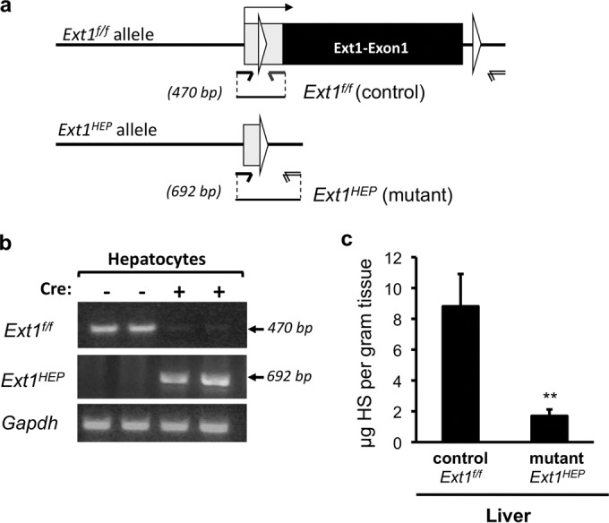
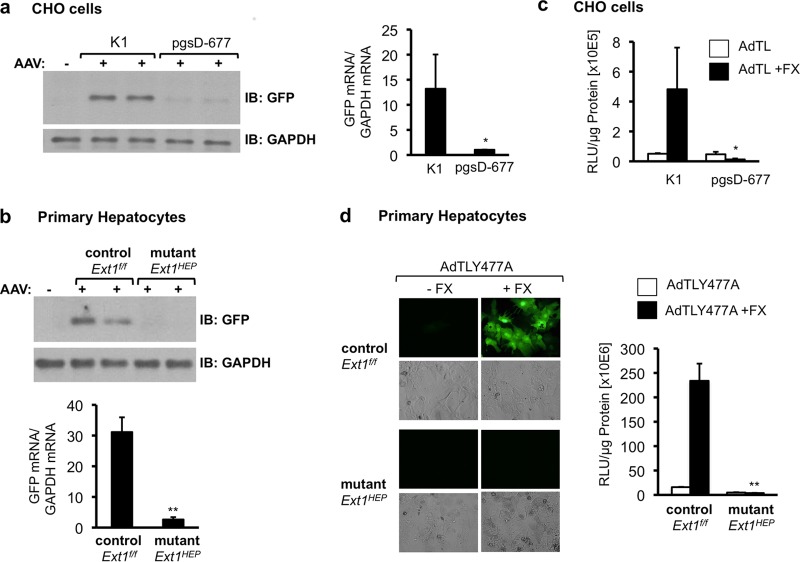
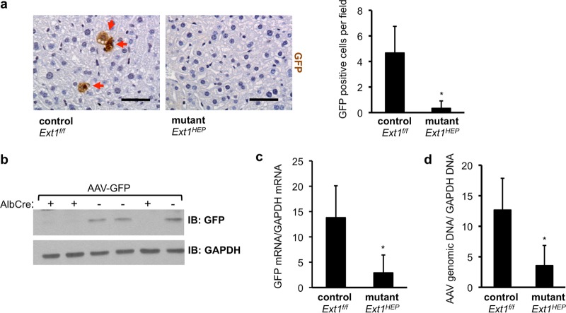
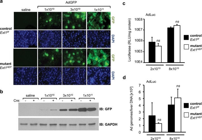
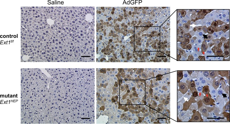
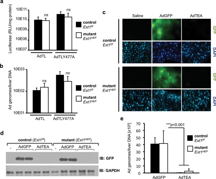
Adeno-associated virus 2 (AAV2) and adenovirus 5 (Ad5) are promising gene therapy vectors. Both display liver tropism and are currently thought to enter hepatocytes in vivo through cell surface heparan sulfate proteoglycans (HSPGs). To test directly this hypothesis, we created mice that lack Ext1, an enzyme required for heparan sulfate biosynthesis, in hepatocytes. Ext1(HEP) mutant mice exhibit an 8-fold reduction of heparan sulfate in primary hepatocytes and a 5-fold reduction of heparan sulfate in whole liver tissue. Conditional hepatocyte Ext1 gene deletion greatly reduced AAV2 liver transduction following intravenous injection. Ad5 transduction requires blood coagulation factor X (FX); FX binds to the Ad5 capsid hexon protein and bridges the virus to HSPGs on the cell surface. Ad5.FX transduction was abrogated in primary hepatocytes from Ext1(HEP) mice. However, in contrast to the case with AAV2, Ad5 transduction was not significantly reduced in the livers of Ext1(HEP) mice. FX remained essential for Ad5 transduction in vivo in Ext1(HEP) mice. We conclude that while AAV2 requires HSPGs for entry into mouse hepatocytes, HSPGs are dispensable for Ad5 hepatocyte transduction in vivo. This study reopens the question of how adenovirus enters cells in vivo.

Importance: Our understanding of how viruses enter cells, and how they can be used as therapeutic vectors to manage disease, begins with identification of the cell surface receptors to which viruses bind and which mediate viral entry. Both adeno-associated virus 2 and adenovirus 5 are currently thought to enter hepatocytes in vivo through heparan sulfate proteoglycans (HSPGs). However, direct evidence for these conclusions is lacking. Experiments presented herein, in which hepatic heparan sulfate synthesis was genetically abolished, demonstrated that HSPGs are not likely to function as hepatocyte Ad5 receptors in vivo. The data also demonstrate that HSPGs are required for hepatocyte transduction by AAV2. These results reopen the question of the identity of the Ad5 receptor in vivo and emphasize the necessity of demonstrating the nature of the receptor by genetic means, both for understanding Ad5 entry into cells in vivo and for optimization of Ad5 vectors as therapeutic agents.


Figures

FIG 1
FIG 1
Ext1HEP mutant mice lack hepatic heparan sulfate. (a) Schematic representation indicating conditional excision of exon 1 of the Ext1 gene via Cre recombinase cleavage at loxP sites (white triangles). Upon Cre recombination, the Ext1 gene is inactivated in hepatocytes (Ext1f/f;AlbCre+ [Ext1HEP]). (b) PCR analysis of Ext1 deletion in primary hepatocytes from Ext1f/f control and Ext1HEP mutant mice, using the primer pairs indicated in panel a. (c) Quantitative glycosaminoglycan analysis in liver tissue. Heparan sulfate (HS) content was analyzed by glycan reductive isotope labeling-LC/MS. The graph illustrates data from 3 control and 3 mutant mice. Values are means ± SD. **, P < 0.01. Comparable results were obtained with two independent preparations of liver tissue.
FIG 2
FIG 2
AAV2 and Ad5 transduction in Ext1-deficient cells in culture. (a) Analysis of GFP transgene expression following AAV transduction of control CHO K1 or EXT1-deficient CHO pgsD-677 cells. Cells were transduced with an AAV2 vector encoding GFP (AAV-GFP) for 1 h in reduced serum-medium. GFP expression was analyzed in cell extracts by immunoblotting (IB) 48 h after transduction (left side). Total RNA was analyzed for GFP mRNA expression by quantitative real-time RT-PCR. GFP mRNA levels are normalized to GAPDH mRNA levels in the same samples (right side). (b) Analysis of GFP protein and RNA expression in primary hepatocyte cultures isolated from Ext1f/f control or Ext1HEP mutant mice 48 h following transduction with AAV-GFP as in panel a. GFP mRNA levels are normalized to mouse GAPDH mRNA levels in the same samples. (c) Transduction of wild-type CHO K1 cells and EXT1-deficient mutant CHO pgsD-677 cells with AdTL, an Ad5 vector carrying a GFP transgene expression cassette and a luciferase transgene expression cassette. The cells were transduced in serum-reduced medium or in serum-reduced medium supplemented with 8 μg/ml of FX. Forty-eight hours posttransduction, cell lysates were analyzed for luciferase transgene expression. (d) Primary cultured hepatocytes from Ext1f/f control or Ext1HEP mutant mice were transduced with AdTLY477A, an Ad5 vector in which CAR binding is ablated, carrying the same GFP and luciferase transgene expression cassettes as AdTL, in the presence or absence of FX. Forty-eight hours following transduction, the cells were analyzed for GFP (by immunofluorescence) and for luciferase transgene expression. RLU, relative light units. Values are means ± SD relative to the value for the control (wild-type CHO K1 cells or control Ext1f/f hepatocytes). *, P < 0.05; **, P < 0.01.
FIG 3
FIG 3
AAV transduction in Ext1f/f control and Ext1HEP mutant mice. Shown is an analysis of liver transduction 8 weeks after intravenous injection of 1 × 10¹¹ VP/mouse of an AAV2 vector encoding green fluorescent protein (AAV-GFP) in Ext1f/f control or Ext1HEP mutant mice. (a) GFP immunohistochemistry in livers from Ext1f/f control or Ext1HEP mutant mice. GFP is visible as brown staining in individual hepatocytes (red arrows). The graph depicts quantification of GFP-positive cells in liver sections. (b) GFP content in liver extracts from Ext1f/f control (Ext1f/f Cre−) or Ext1HEP mutant (Ext1f/f Cre+) mice. Liver extracts were assayed for GFP and GAPDH by immunoblotting. “+” indicates the presence of the AlbCre transgene, leading to the targeted deletion of the Ext1 gene to create Ext1HEP mutant mice; “−” indicates the absence of the AlbCre transgene, resulting in Ext1f/f mice with an intact Ext1 gene. (c) Total liver RNA was analyzed for GFP mRNA expression by quantitative real-time RT-PCR and normalized to the mouse liver GAPDH mRNA of the same samples. (d) AAV vector genomes in livers from AAV-injected Ext1f/f control or Ext1HEP mutant mice were quantified after DNA extraction by quantitative real-time PCR, using primers within the CMV promoter of the transgene expression cassette, and normalized to liver GAPDH DNA values of the same sample. Values are means ± SD. Differences in number of GFP-expressing cells per field, GFP mRNA expression, and vector genomes between control and mutant mice were compared by Student's t test. *, P < 0.05; n = 3. Scale bar represents 50 μm.
FIG 4
FIG 4
Adenovirus transduction in Ext1f/f control and Ext1HEP mutant mice. (a) GFP transgene expression in livers of Ext1f/f control or mutant Ext1HEP mice 3 days after intravenous injection with increasing doses (1 × 10¹⁰, 3 × 10¹⁰, or 1 × 10¹¹VP/mouse) of an Ad5 vector expressing GFP. GFP content was visualized by immunofluorescence staining of paraffin-embedded liver sections. Corresponding fields stained with DAPI are displayed to visualize nuclei. (b) Liver extracts were analyzed by immunoblotting for GFP and GAPDH. Liver extracts from two representative mice per group are shown. (c and d) Liver transduction using a low dose (2 × 10¹⁰ VP/mouse) and a high dose (8 × 10¹⁰ VP/mouse) of AdLuc, an Ad5 vector encoding firefly luciferase. Luciferase transgene expression (c) and adenovirus vector genomes (d) in Ext1f/f control and Ext1HEP mutant mice were analyzed 3 days after intravenous injection. Ad5 genomes were detected in liver by quantitative PCR. Values are means ± SD. Differences in either transgene expression or vector genomes between control and mutant mice were compared by Student's t test. ns, not significant (P > 0.05); n = 4 mice per group.
FIG 5
FIG 5
Histological analysis of liver sections from adenovirus-transduced Ext1f/f control and Ext1HEP mutant mice. Immunohistochemistry results for GFP in liver sections from Ext1f/f control and Ext1HEP mutant mice 3 days following intravenous injection with 8 × 10¹⁰ AdGFP VP/mouse are shown. GFP in cells is visible as dark-brown staining throughout the cytoplasm. Red arrows indicate stained (GFP-positive) and unstained (GFP-negative) cells side by side. Hematoxylin counterstaining was used to visualize nuclei in blue (black arrows). White arrows indicate sinusoids and bile ducts. Scale bar indicates 50 μm.
FIG 6
FIG 6
The roles of CAR binding and FX binding in Ad5 liver transduction in Ext1f/f control and Ext1HEP mutant mice. Shown is luciferase transgene expression (a) and viral genomes (b) in the livers of Ext1f/f control and Ext1HEP mutant mice 3 days after intravenous injection of 5 × 10¹⁰ VP/mouse of AdTLY477A, a luciferase-GFP-expressing Ad5 vector in which CAR binding is ablated, or the corresponding unmodified AdTL control vector. (c) Histological assessment of AdGFP- and AdTEA-mediated GFP expression in frozen sections of Ext1f/f control and Ext1HEP mutant mouse livers 3 days after intravenous injection with 1.5 × 10¹¹ viral particles/mouse. AdTEA is a GFP-expressing Ad5 vector in which FX binding is ablated. DAPI-stained fields are displayed to visualize nuclei. (d) Immunoblot analysis of GFP expression in the livers of Ext1f/f and Ext1HEP mice injected with AdGFP or AdTEA adenovirus vectors. GFP expression in liver extracts from two representative mice per group is shown. (e) Viral genome content in liver tissue of Ext1f/f and Ext1HEP mice injected with AdGFP or AdTEA. DNA was extracted from the same livers as in panels c and d, and viral genomes were quantified using quantitative real-time PCR. Individual groups were compared by Student's t test. Values are displayed as means ± SD. **, P < 0.01; ***, P < 0.001. n = 4.

Similar articles

Cited by

References

    1. Mingozzi F, High KA. 2011. Therapeutic in vivo gene transfer for genetic disease using AAV: progress and challenges. Nat Rev Genet 12:341–355. doi:10.1038/nrg2988. - DOI - PubMed
    1. Nienhuis AW. 2013. Development of gene therapy for blood disorders: an update. Blood 122:1556–1564. doi:10.1182/blood-2013-04-453209. - DOI - PMC - PubMed
    1. Kern A, Schmidt K, Leder C, Muller OJ, Wobus CE, Bettinger K, Von der Lieth CW, King JA, Kleinschmidt JA. 2003. Identification of a heparin-binding motif on adeno-associated virus type 2 capsids. J Virol 77:11072–11081. doi:10.1128/JVI.77.20.11072-11081.2003. - DOI - PMC - PubMed
    1. Koeberl DD, Alexander IE, Halbert CL, Russell DW, Miller AD. 1997. Persistent expression of human clotting factor IX from mouse liver after intravenous injection of adeno-associated virus vectors. Proc Natl Acad Sci U S A 94:1426–1431. doi:10.1073/pnas.94.4.1426. - DOI - PMC - PubMed
    1. Tomko RP, Xu R, Philipson L. 1997. HCAR and MCAR: the human and mouse cellular receptors for subgroup C adenoviruses and group B coxsackieviruses. Proc Natl Acad Sci U S A 94:3352–3356. doi:10.1073/pnas.94.7.3352. - DOI - PMC - PubMed

Publication types