. 2015 Oct 21;90(1):521-32.
doi: 10.1128/JVI.02014-15. Print 2016 Jan 1.

A Single Maturation Cleavage Site in Adenovirus Impacts Cell Entry and Capsid Assembly

Affiliations

A Single Maturation Cleavage Site in Adenovirus Impacts Cell Entry and Capsid Assembly

Crystal L Moyer et al. J Virol. .

Abstract

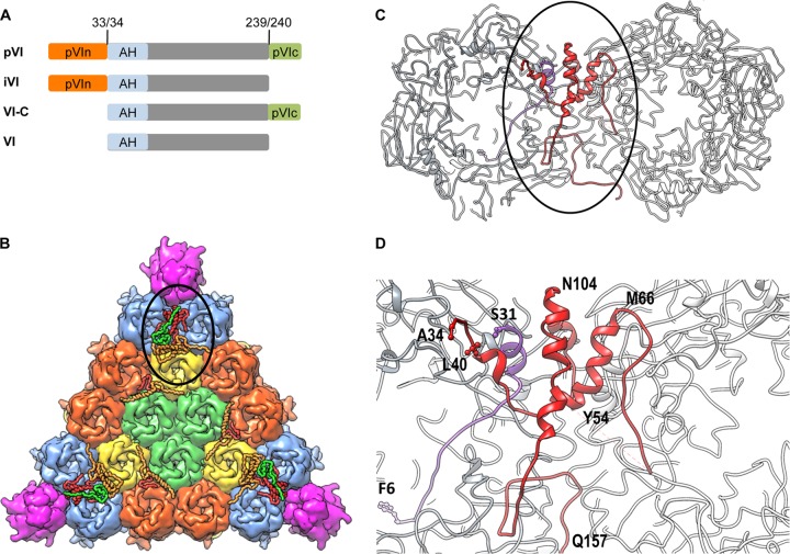
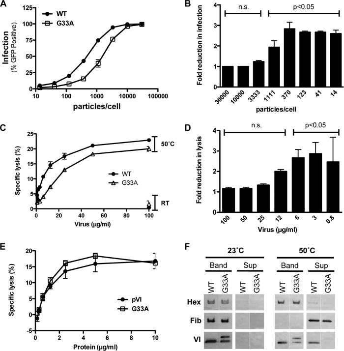
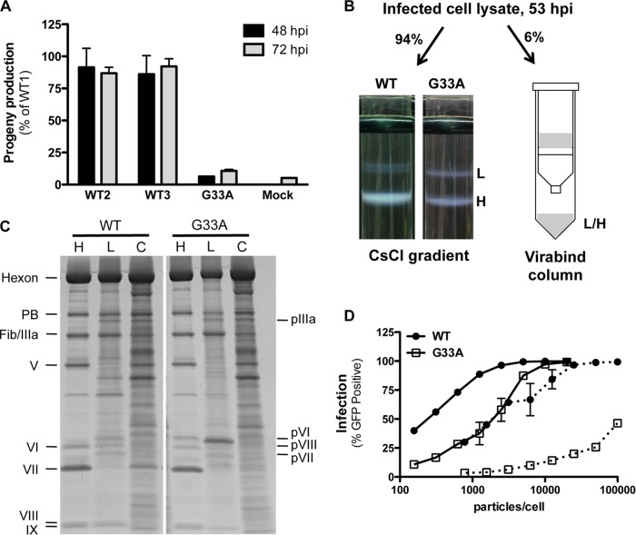
Proteolytic maturation drives the conversion of stable, immature virus particles to a mature, metastable state primed for cell infection. In the case of human adenovirus, this proteolytic cleavage is mediated by the virally encoded protease AVP. Protein VI, an internal capsid cement protein and substrate for AVP, is cleaved at two sites, one of which is near the N terminus of the protein. In mature capsids, the 33 residues at the N terminus of protein VI (pVIn) are sequestered inside the cavity formed by peripentonal hexon trimers at the 5-fold vertex. Here, we describe a glycine-to-alanine mutation in the N-terminal cleavage site of protein VI that profoundly impacts proteolytic processing, the generation of infectious particles, and cell entry. The phenotypic effects associated with this mutant provide a mechanistic framework for understanding the multifunctional nature of protein VI. Based on our findings, we propose that the primary function of the pVIn peptide is to mediate interactions between protein VI and hexon during virus replication, driving hexon nuclear accumulation and particle assembly. Once particles are assembled, AVP-mediated cleavage facilitates the release of the membrane lytic region at the amino terminus of mature VI, allowing it to lyse the endosome during cell infection. These findings highlight the importance of a single maturation cleavage site for both infectious particle production and cell entry and emphasize the exquisite spatiotemporal regulation governing adenovirus assembly and disassembly.

Importance: Postassembly virus maturation is a cornerstone principle in virology. However, a mechanistic understanding of how icosahedral viruses utilize this process to transform immature capsids into infection-competent particles is largely lacking. Adenovirus maturation involves proteolytic processing of seven precursor proteins. There is currently no information for the role of each independent cleavage event in the generation of infectious virions. To address this, we investigated the proteolytic maturation of one adenovirus precursor molecule, protein VI. Structurally, protein VI cements the outer capsid shell and links it to the viral core. Functionally, protein VI is involved in endosome disruption, subcellular trafficking, transcription activation, and virus assembly. Our studies demonstrate that the multifunctional nature of protein VI is largely linked to its maturation. Through mutational analysis, we show that disrupting the N-terminal cleavage of preprotein VI has major deleterious effects on the assembly of infectious virions and their subsequent ability to infect host cells.


Figures

FIG 1
FIG 1
Protein VI maturation and location within AdV particles. (A) Schematic of protein VI. Preprotein VI (pVI) is cleaved by the adenoviral cysteine protease (AVP) at both termini (numbered residues) to generate mature protein VI (gray). Two intermediate species cleaved at one of the two termini (iVI and VI-C) are also illustrated. The N-terminal pVIn peptide (orange) binds the internal cavity of hexon trimers. The pVIc peptide (green) is a cofactor for AVP. The membrane lytic amphipathic helix (AH), present at the N terminus of mature VI, is highlighted in light blue. (B) Crystal structure of one interior facet of the HAdV5 capsid. Color schemes are as follows: hexons, blue, orange, yellow, and light green; penton base, magenta; protein VI, red; protein V, green; protein VIII, light orange. The location of the complex formed by proteins VI, V, and VIII beneath the peripentonal hexons is circled. (C) Zoom-in view of the circled area from panel B, with the peripentonal hexons shown in gray, pVIn shown in purple, and mature VI shown in red. (D) Zoom-in view of the circled area from panel C, using the same color scheme. Selected residues from pVIn and mature VI are designated using the single-amino-acid code. PDB identifier, 4CWU.
FIG 2
FIG 2
Preprotein VI, but not mature VI, associates with hexon. (A) Hexon binding by recombinant protein VI. ELISA plates, coated with recombinant pVI or VI, were incubated with serial dilutions of hexon, and then hexon was detected with an antihexon antibody. (B) Competition ELISA between different recombinant protein VI molecules and pVIn peptide. ELISA plates coated with pVI or VI were incubated with hexon, hexon preincubated with a 100-fold molar excess of pVIn, or buffer. The pVIn peptide competes only with pVI for hexon binding. An unpaired two-tailed t test was used to calculate statistical significance. (C) The membrane lytic activities of pVI and VI are similar. Serial dilutions of recombinant protein VI were incubated with SulfoB-entrapped liposomes, and specific lysis was measured using a Fluorimager. (D) Hexon attenuates the membrane lytic activity of pVI but not VI. pVI or VI (150 nM) was mixed with serial dilutions of hexon and then added to liposomes. Data are the percentage of specific lysis compared to the lytic activity of pVI or VI in the absence of hexon. P values were calculated using a one-way analysis of variance and Dunnett's multiple-comparison test (compared to the 0 nM hexon control). n.s., not significant (P > 0.05).
FIG 3
FIG 3
A single G33A mutation in the N-terminal AVP cleavage site of protein VI reduces in vitro processing. (A) Amino acid sequence alignment of the N-terminal regions of protein VI in different human AdVs. The consensus sequence for recognition by AVP is given at the top of the table. Directly below is an alignment of the pVI N-terminal cleavage site sequences for different species (A to G). The HAdV5 protein VI cleavage site sequence spans residues 30 to 34. The highly conserved glycine residue at position 33 was mutated to alanine, and the mutation is indicated by an asterisk. (B) In vitro cleavage of pVI by AVP. Recombinant pVI-Strep was incubated with recombinant AVP for the indicated number of minutes at room temperature. Labeled bands to the left of the gel correspond with the labeling scheme in the schematic below, which describes the different recombinant proteins and indicates the expected molecular mass of each following processing at the N and/or C terminus by AVP. A nonspecific degradation product copurifies with the full-length proteins (slightly lower band at the 0-min point).
FIG 4
FIG 4
The G33A mutation impacts virion composition and pVI cleavage in infected cells. (A) SDS-PAGE gel analysis of purified virus particles, including Ad5, Ad5-Pro-P137L (produces immature virions harboring precursor proteins), and Ad5-pVI-G33A. The gel in the upper panel was stained with Simply Blue and reveals various proteins from mature and immature HAdV5 that are labeled as indicated. The lower panel shows an immunoblot assay for protein VI. The additional protein VI band in the G33A virus is iVI, as it migrates differently (in between) than both pVI and mature VI. (B) Two independent preparations of wild-type (WT) or G33A virions (labeled 1 and 2) were analyzed by SDS-PAGE and SYPRO Ruby staining. Numbers at left are molecular masses in kilodaltons. (C) Quantities of the total amount of protein VI incorporated into virus particles determined by densitometry analysis of the gels shown in panel B and normalized to each of the other virus structural proteins as described in Materials and Methods. The data are the mean percentage ± SEM of protein VI incorporation shown for all viral proteins, compared to wild-type preparation 1. (D) Time course of pVI cleavage in infected cells. Lysates of infected cells were made at the indicated times (hpi) and immunoblotted for protein VI. Note the significant overall reduction and delayed processing of pVI to mature VI for the G33A point mutant.
FIG 5
FIG 5
The G33A mutation attenuates infection and membrane lysis. (A) Single-round infection. Noncomplementing (E1-negative) A549 cells were infected with the indicated virus for 48 h, and the percentage of infected (GFP+) cells was determined by flow cytometry. WT, wild type. (B) Fold reduction in G33A mutant virus infection as a function of input particles/cell compared to wild-type virus. The data are derived from the samples depicted in panel A. P values are calculated with a one-way analysis of variance and Dunnett's multiple-comparison test. n.s., not significant. (C) Liposome lysis by heat-disrupted virus. Various amounts of viruses were incubated at room temperature (RT) (control) or heated to 50°C for 10 min to liberate protein VI and then mixed with liposomes. The percent specific lysis for each concentration is indicated. (D) Fold reduction in liposome lysis as a function of virus concentration compared to wild-type virus. The data are a subset of samples depicted in panel C. P values are calculated with a one-way analysis of variance and Dunnett's multiple-comparison test. (E) Liposome lysis by recombinant protein VI. Serial dilutions of wild-type and G33A mutant proteins were incubated with fluorescent dye-entrapped liposomes, and specific lysis was measured with a Fluorimager. (F) Thermostability assay to compare the dissociations of capsid proteins in mutant and wild-type viruses. Wild-type or mutant viruses were incubated at the indicated temperature and then subjected to density gradient ultracentrifugation to separate core (Band) and released (Sup) proteins. Proteins (hexon, fiber, and VI) were visualized via immunoblotting with the indicated antibodies.
FIG 6
FIG 6
The G33A cleavage site mutant reduces infectious particle assembly. (A) Virus burst assay. Complementing 293β5 (E1-positive) cells were infected with equivalent amounts of infectious particles in triplicate. Cell lysates were prepared 48 and 72 hpi and passaged onto noncomplementing A549 cells. Virus infection (GFP transgene expression) was measured 48 hpi. Three independent preparations of wild-type (WT) Ad5 were included to assess intrinsic variations in virus burst. Progeny production is shown as a percentage of the value for wild-type preparation 1. (B) Illustration of sample preparation for studies shown in panels C and D. Infected cell lysates were divided as indicated, and virus was purified by cesium chloride density gradient ultracentrifugation or by binding and elution in a Virabind column. The light (L, genome-lacking) and heavy (H, genome-containing) virus bands were harvested separately from cesium chloride gradients, while the eluate from the Virabind column contains both heavy and light particles. (C) SDS-PAGE analysis of the virus purified by the different methods described in the legend to panel B. Note the differences in precursor capsid protein processing in the heavy and light particles. H, heavy; L, light; C, column purified. (D) The G33A cleavage mutant severely impacts particle assembly. Equivalent numbers of particles from the samples generated in panel B were used to infect A549 cells, and infection (GFP expression) was measured by flow cytometry. Mature viruses from the cesium chloride gradient are shown as solid lines, while column-purified virus is indicated with dotted lines.

Similar articles

Cited by

References

    1. Mangel WF, San Martin C. 2014. Structure, function and dynamics in adenovirus maturation. Viruses 6:4536–4570. doi:10.3390/v6114536. - DOI - PMC - PubMed
    1. Webster A, Russell S, Talbot P, Russell WC, Kemp GD. 1989. Characterization of the adenovirus proteinase: substrate specificity. J Gen Virol 70:3225–3234. doi:10.1099/0022-1317-70-12-3225. - DOI - PubMed
    1. Weber J. 1976. Genetic analysis of adenovirus type 2 III. Temperature sensitivity of processing viral proteins. J Virol 17:462–471. - PMC - PubMed
    1. Anderson CW. 1990. The proteinase polypeptide of adenovirus serotype 2 virions. Virology 177:259–272. doi:10.1016/0042-6822(90)90479-B. - DOI - PubMed
    1. Rancourt C, Keyvani-Amineh H, Sircar S, Labrecque P, Weber JM. 1995. Proline 137 is critical for adenovirus protease encapsidation and activation but not enzyme activity. Virology 209:167–173. doi:10.1006/viro.1995.1240. - DOI - PubMed

Publication types

Associated data

LinkOut - more resources