Review
. 2015:2015:795602.
doi: 10.1155/2015/795602. Epub 2015 Sep 29.

Chemotherapy-Induced Cardiotoxicity: Overview of the Roles of Oxidative Stress

Affiliations
Review

Chemotherapy-Induced Cardiotoxicity: Overview of the Roles of Oxidative Stress

Paweorn Angsutararux et al. Oxid Med Cell Longev. 2015.

Abstract

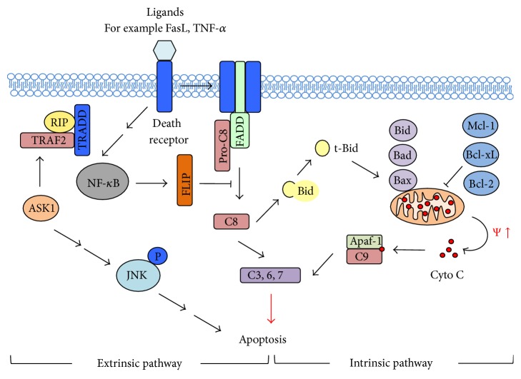
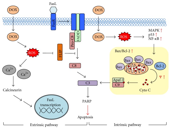
Chemotherapy-induced cardiotoxicity is a serious complication that poses a serious threat to life and limits the clinical use of various chemotherapeutic agents, particularly the anthracyclines. Understanding molecular mechanisms of chemotherapy-induced cardiotoxicity is a key to effective preventive strategies and improved chemotherapy regimen. Although no reliable and effective preventive treatment has become available, numerous evidence demonstrates that chemotherapy-induced cardiotoxicity involves the generation of reactive oxygen species (ROS). This review provides an overview of the roles of oxidative stress in chemotherapy-induced cardiotoxicity using doxorubicin, which is one of the most effective chemotherapeutic agents against a wide range of cancers, as an example. Current understanding in the molecular mechanisms of ROS-mediated cardiotoxicity will be explored and discussed, with emphasis on cardiomyocyte apoptosis leading to cardiomyopathy. The review will conclude with perspectives on model development needed to facilitate further progress and understanding on chemotherapy-induced cardiotoxicity.


Figures

Figure 1
Figure 1
Major pathways of apoptosis. An extrinsic pathway of apoptosis (left) involves the stimulation through binding of death receptor (e.g., FasR and TNFR) to their respective ligands (e.g., FasL and TNF-α), recruiting adaptive proteins such as Fas-associated death-domain (FADD) and pro-caspase-8 (pro-C8), forming death-inducing signaling complex (DISC) and relaying signals to activation of effector caspases such as caspase-3 (C3), C6, and C7. In addition, Bid is also activated, which transduces these death signals to the intrinsic pathway. On the other hand, an intrinsic pathway of apoptosis is induced in response to cellular stresses such as DNA damage and ROS that increase the expression of proapoptotic Bcl-2 family proteins (e.g., Bid, Bad, and Bax), while repressing antiapoptotic Bcl-2 family proteins (e.g., Bcl-2, Bcl-xL, and Mcl-1), leading to an alteration in mitochondrial membrane potential and the release of cytochrome C (Cyto C). Proteins such as Apaf-1 and caspase-9 (C9) are activated, resulting in the formation of an apoptosome, which then stimulate the activation of effector caspases. NF-κB and JNK/ASK-1 also play a role in apoptotic signaling through the regulation of antiapoptotic molecules such as FLIP and Bcl-2.
Figure 2
Figure 2
Schematic representation of DOX-induced apoptosis and the involvement of ROS. DOX-derived ROS could affect the regulation of calcium homeostasis, resulting in cytosolic calcium (Ca2+) overload that can activate calcineurin and increase the transcription of Fas ligand (FasL). DOX-derived ROS could also inhibit the expression of caspase-8 (C8, also known as FLICE) inhibitory protein FLIP, rendering cells to apoptosis. Additionally, DOX-derived ROS could act as an intrinsic stress that activates mitogen activated protein kinases (MAPK) p38 and JNK and NF-κB pathways as well as intracellular p53 accumulation, leading to an alteration in the ratio of proapoptotic proteins to antiapoptotic proteins (e.g., Bax to Bcl-2), cytochrome C (Cyto C) release, and caspase-9 and -3 (C9/C3) activation.

References

    1. Kufe D. W., Holland J. F., Frei E. American Cancer Society: Cancer Medicine. Hamilton, Canada: BC Decker; 2003.
    1. Hershman D. L., McBride R. B., Eisenberger A., Wei Y. T., Grann V. R., Jacobson J. S. Doxorubicin, cardiac risk factors, and cardiac toxicity in elderly patients with diffuse B-cell non-Hodgkin's lymphoma. Journal of Clinical Oncology. 2008;26(19):3159–3165. doi: 10.1200/jco.2007.14.1242. - DOI - PubMed
    1. Gianni L., Munzone E., Capri G., et al. Paclitaxel by 3-hour infusion in combination with bolus doxorubicin in women with untreated metastatic breast cancer: high antitumor efficacy and cardiac effects in a dose-finding and sequence-finding study. Journal of Clinical Oncology. 1995;13(11):2688–2699. - PubMed
    1. Eisenhauer E. A., Vermorken J. B. The taxoids. Comparative clinical pharmacology and therapeutic potential. Drugs. 1998;55(1):5–30. doi: 10.2165/00003495-199855010-00002. - DOI - PubMed
    1. Giantris A., Abdurrahman L., Hinkle A., Asselin B., Lipshultz S. E. Anthracycline-induced cardiotoxicity in children and young adults. Critical Reviews in Oncology/Hematology. 1998;27(1):53–68. doi: 10.1016/S1040-8428(97)10007-5. - DOI - PubMed

Publication types

Substances