

Enamel Pearls Implications on Periodontal Disease
- PMID: 26491574
- PMCID: PMC4600495
- DOI: 10.1155/2015/236462
Enamel Pearls Implications on Periodontal Disease
Abstract
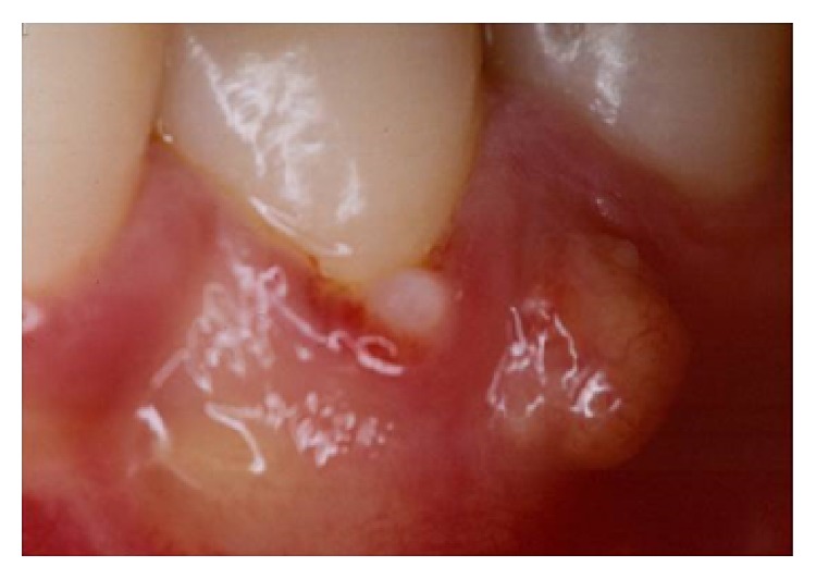
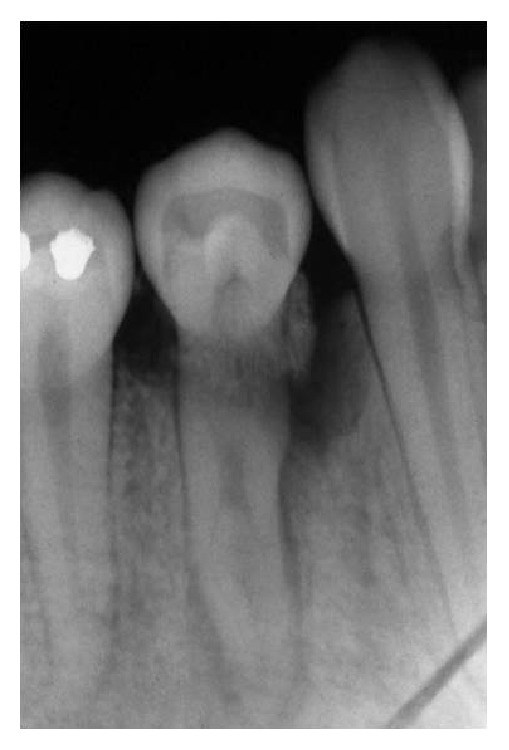
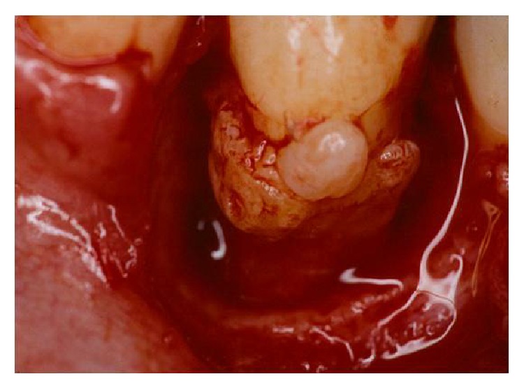
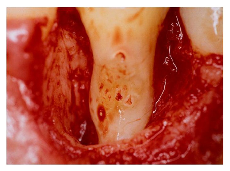
Dental anatomy is quite complex and diverse factors must be taken into account in its analysis. Teeth with anatomical variations present an increase in the rate of severity periodontal tissue destruction and therefore a higher risk of developing periodontal disease. In this context, this paper reviews the literature regarding enamel pearls and their implications in the development of severe localized periodontal disease as well as in the prognosis of periodontal therapy. Radiographic examination of a patient complaining of pain in the right side of the mandible revealed the presence of a radiopaque structure around the cervical region of lower right first premolar. Periodontal examination revealed extensive bone loss since probing depths ranged from 7.0 mm to 9.0 mm and additionally intense bleeding and suppuration. Surgical exploration detected the presence of an enamel pearl, which was removed. Assessment of the remaining supporting tissues led to the extraction of tooth 44. Local factors such as enamel pearls can lead to inadequate removal of the subgingival biofilm, thus favoring the establishment and progression of periodontal diseases.
Figures
Similar articles
-
Treatment of an Unusual Non-Tooth Related Enamel Pearl (EP) and 3 Teeth-Related EPs with Localized Periodontal Disease Without Teeth Extractions: A Case Report.Compend Contin Educ Dent. 2015 Sep;36(8):592-9. Compend Contin Educ Dent. 2015. PMID: 26355443
-
Prevalence of enamel pearls in teeth from a human teeth bank.J Oral Sci. 2010 Jun;52(2):257-60. doi: 10.2334/josnusd.52.257. J Oral Sci. 2010. PMID: 20587950
-
Enamel pearl on an unusual location associated with localized periodontal disease: A clinical report.J Indian Soc Periodontol. 2013 Nov;17(6):796-800. doi: 10.4103/0972-124X.124520. J Indian Soc Periodontol. 2013. PMID: 24554894 Free PMC article.
-
Enamel pearls and cervical enamel projections on 2 maxillary molars with localized periodontal disease: case report and histologic study.Oral Surg Oral Med Oral Pathol Oral Radiol Endod. 2000 Apr;89(4):493-7. doi: 10.1016/s1079-2104(00)70131-4. Oral Surg Oral Med Oral Pathol Oral Radiol Endod. 2000. PMID: 10760733 Review.
-
Detection of localized tooth-related factors that predispose to periodontal infections.Periodontol 2000. 2004;34:136-50. doi: 10.1046/j.0906-6713.2003.003429.x. Periodontol 2000. 2004. PMID: 14717860 Review.
Cited by
-
Furcation Involvement in Periodontal Disease: A Narrative Review.Cureus. 2024 Mar 10;16(3):e55924. doi: 10.7759/cureus.55924. eCollection 2024 Mar. Cureus. 2024. PMID: 38601385 Free PMC article. Review.
-
Biometric analysis of furcation area of molar teeth and its relationship with instrumentation.BMC Oral Health. 2024 Apr 10;24(1):436. doi: 10.1186/s12903-024-04164-2. BMC Oral Health. 2024. PMID: 38600486 Free PMC article.
-
Developmental Structural Tooth Defects in Dogs - Experience From Veterinary Dental Referral Practice and Review of the Literature.Front Vet Sci. 2016 Feb 8;3:9. doi: 10.3389/fvets.2016.00009. eCollection 2016. Front Vet Sci. 2016. PMID: 26904551 Free PMC article. Review.
References
-
- Löe H., Theiland E., Jensen S. B. Experimental gingivitis in man. Journal of Periodontology. 1965;36:177–186. - PubMed
-
- Gher M. E., Vernino A. R. Root anatomy: a local factor in inflammatory periodontal disease. The International Journal of Periodontics & Restorative Dentistry. 1981;1(5):53–63. - PubMed
LinkOut - more resources
Full Text Sources
Other Literature Sources
