

Ultrastructural Changes in Clinical and Microbiota Isolates of Klebsiella pneumoniae Carriers of Genes bla SHV, bla TEM, bla CTX-M, or bla KPC When Subject to β-Lactam Antibiotics
- PMID: 26491715
- PMCID: PMC4600873
- DOI: 10.1155/2015/572128
Ultrastructural Changes in Clinical and Microbiota Isolates of Klebsiella pneumoniae Carriers of Genes bla SHV, bla TEM, bla CTX-M, or bla KPC When Subject to β-Lactam Antibiotics
Abstract
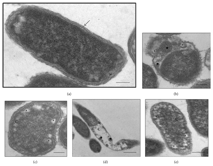
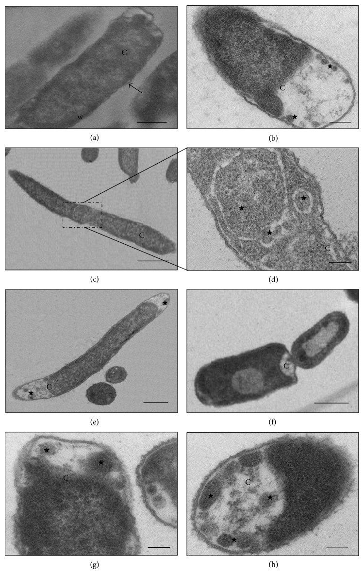
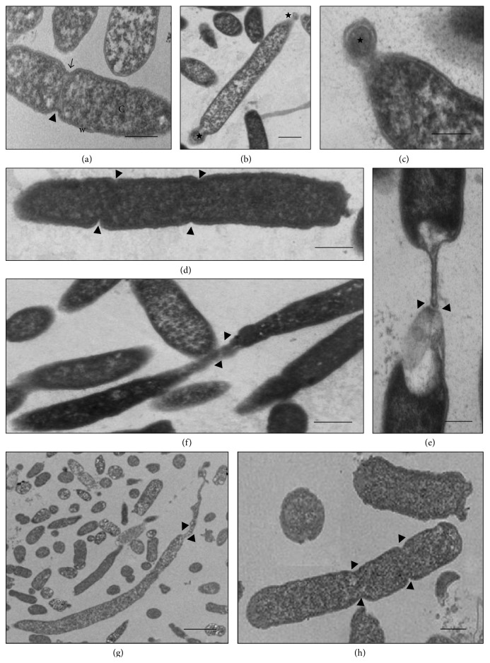
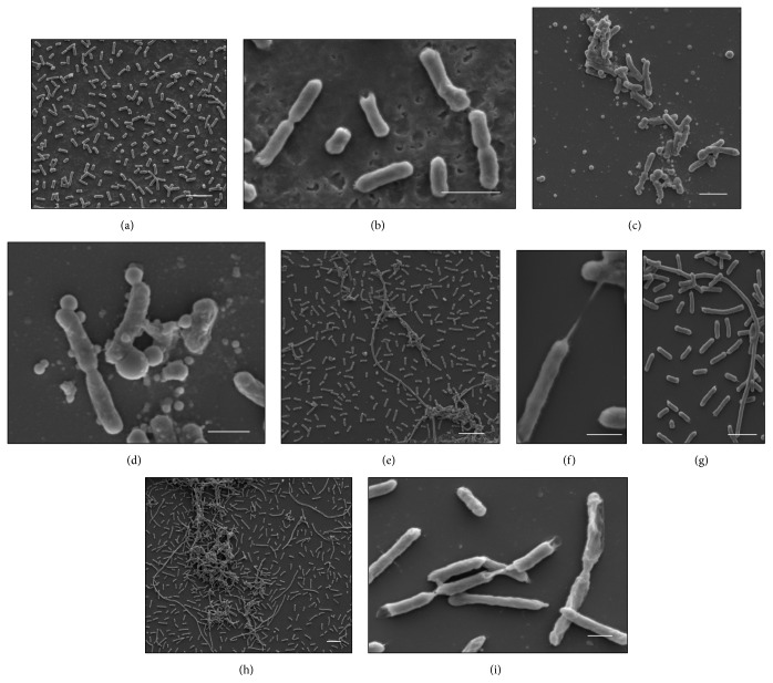
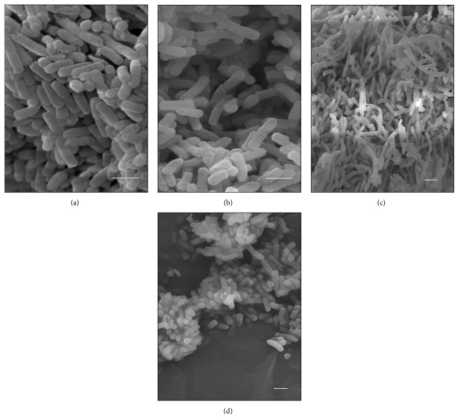
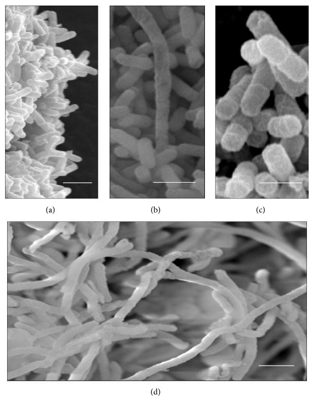
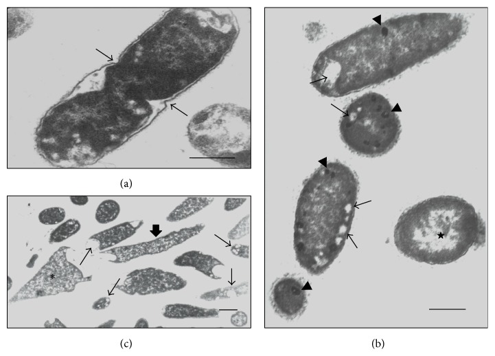
The aim of this study was to characterize the ultrastructural effects caused by β-lactam antibiotics in Klebsiella pneumoniae isolates. Three K. pneumoniae clinical isolates were selected for the study with resistance profiles for third-generation cephalosporins, aztreonam, and/or imipenem and with different resistance genes for extended-spectrum β-lactamases (ESBL) or Klebsiella pneumoniae carbapenemase (KPC). Two K. pneumoniae isolates obtained from the microbiota, which were both resistant to amoxicillin and ampicillin, were also analyzed. In accordance with the susceptibility profile, the clinical isolates were subjected to subminimum inhibitory concentrations (sub-MICs) of cefotaxime, ceftazidime, aztreonam, and imipenem and the isolates from the microbiota to ampicillin and amoxicillin, for analysis by means of scanning and transmission electron microscopy. The K. pneumoniae isolates showed different morphological and ultrastructural changes after subjection to β-lactams tested at different concentrations, such as cell filamentation, loss of cytoplasmic material, and deformation of dividing septa. Our results demonstrate that K. pneumoniae isolates harboring different genes that encode for β-lactamases show cell alterations when subjected to different β-lactam antibiotics, thus suggesting that they possess residual activity in vitro, despite the phenotypic resistance presented in the isolates analyzed.
Figures
Similar articles
-
Distribution of bla(TEM), bla(SHV), bla(CTX-M) genes among clinical isolates of Klebsiella pneumoniae at Labbafinejad Hospital, Tehran, Iran.Microb Drug Resist. 2010 Mar;16(1):49-53. doi: 10.1089/mdr.2009.0096. Microb Drug Resist. 2010. PMID: 19961397
-
Phenotypic and genotypic characterization of β-lactam resistance in Klebsiella pneumoniae isolated from swine.Vet Microbiol. 2011 Apr 21;149(1-2):139-46. doi: 10.1016/j.vetmic.2010.09.030. Epub 2010 Oct 7. Vet Microbiol. 2011. PMID: 21035968
-
Carbapenem-hydrolysing beta-lactamase KPC-2 in Klebsiella pneumoniae isolated in Rio de Janeiro, Brazil.J Antimicrob Chemother. 2009 Feb;63(2):265-8. doi: 10.1093/jac/dkn484. Epub 2008 Nov 20. J Antimicrob Chemother. 2009. PMID: 19028717
-
Underlying mechanisms of carbapenem resistance in extended-spectrum β-lactamase-producing Klebsiella pneumoniae and Escherichia coli isolates at a tertiary care centre in Lebanon: role of OXA-48 and NDM-1 carbapenemases.Int J Antimicrob Agents. 2013 Jan;41(1):75-9. doi: 10.1016/j.ijantimicag.2012.08.010. Epub 2012 Nov 9. Int J Antimicrob Agents. 2013. PMID: 23142087
-
Identification and characterization of CTX-M-15 producing Klebsiella pneumoniae clone ST101 in a Hungarian university teaching hospital.Acta Microbiol Immunol Hung. 2015 Sep;62(3):233-45. doi: 10.1556/030.62.2015.3.2. Acta Microbiol Immunol Hung. 2015. PMID: 26551567 Review.
Cited by
-
Soil Fungi as Biomediator in Silver Nanoparticles Formation and Antimicrobial Efficacy.Int J Nanomedicine. 2022 Jun 29;17:2843-2863. doi: 10.2147/IJN.S356724. eCollection 2022. Int J Nanomedicine. 2022. PMID: 35795079 Free PMC article.
-
Designing phage cocktails to combat the emergence of bacteriophage-resistant mutants in multidrug-resistant Klebsiella pneumoniae.Microbiol Spectr. 2024 Jan 11;12(1):e0125823. doi: 10.1128/spectrum.01258-23. Epub 2023 Nov 29. Microbiol Spectr. 2024. PMID: 38018985 Free PMC article.
-
Molecular Nanomachines Disrupt Bacterial Cell Wall, Increasing Sensitivity of Extensively Drug-Resistant Klebsiella pneumoniae to Meropenem.ACS Nano. 2019 Dec 24;13(12):14377-14387. doi: 10.1021/acsnano.9b07836. Epub 2019 Dec 11. ACS Nano. 2019. PMID: 31815423 Free PMC article.
References
Publication types
MeSH terms
Substances
LinkOut - more resources
Full Text Sources
Other Literature Sources
Medical
