. 2016 Jan;80(1):16-23; discussion 23-5.
doi: 10.1097/TA.0000000000000885.

Overwhelming tPA release, not PAI-1 degradation, is responsible for hyperfibrinolysis in severely injured trauma patients

Affiliations

Overwhelming tPA release, not PAI-1 degradation, is responsible for hyperfibrinolysis in severely injured trauma patients

Michael P Chapman et al. J Trauma Acute Care Surg. 2016 Jan.

Abstract

Background: Trauma-induced coagulopathy (TIC) is associated with a fourfold increased risk of mortality. Hyperfibrinolysis is a component of TIC, but its mechanism is poorly understood. Plasminogen activation inhibitor (PAI-1) degradation by activated protein C has been proposed as a mechanism for deregulation of the plasmin system in hemorrhagic shock, but in other settings of ischemia, tissue plasminogen activator (tPA) has been shown to be elevated. We hypothesized that the hyperfibrinolysis in TIC is not the result of PAI-1 degradation but is driven by an increase in tPA, with resultant loss of PAI-1 activity through complexation with tPA.

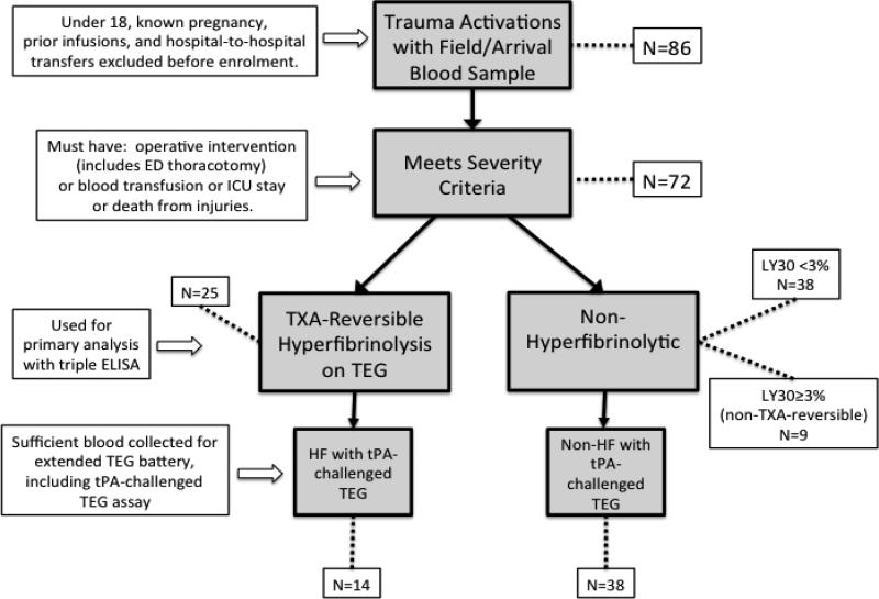
Methods: Eighty-six consecutive trauma activation patients had blood collected at the earliest time after injury and were screened for hyperfibrinolysis using thrombelastography (TEG). Twenty-five hyperfibrinolytic patients were compared with 14 healthy controls using enzyme-linked immunosorbent assays for active tPA, active PAI-1, and PAI-1/tPA complex. Blood was also subjected to TEG with exogenous tPA challenge as a functional assay for PAI-1 reserve.

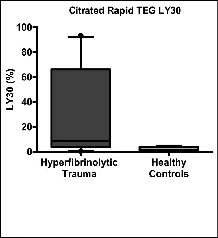
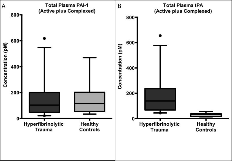
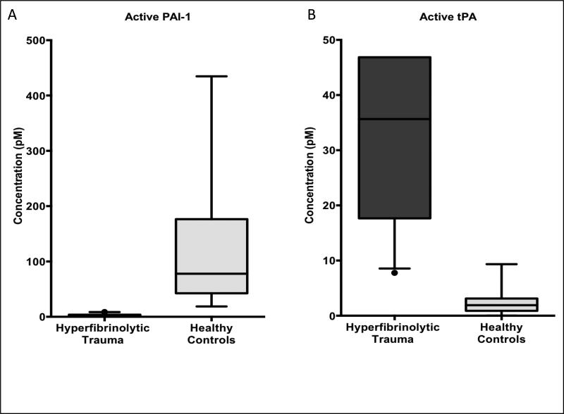
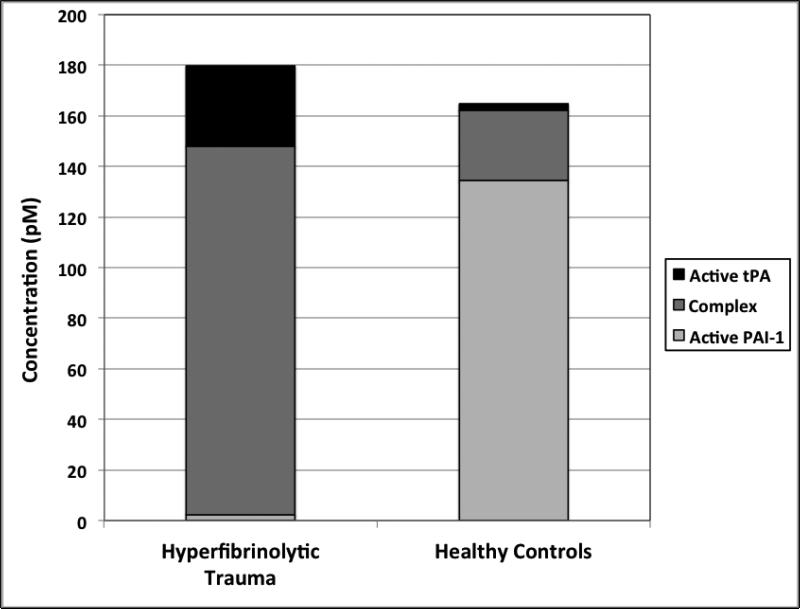
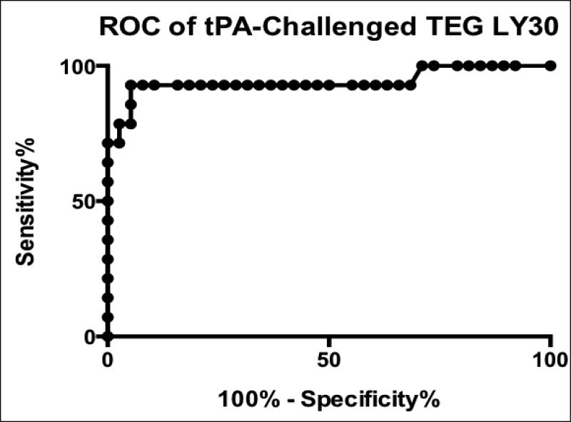
Results: Total levels of PAI-1 (the sum of the active PAI-1 species and its covalent complex with tPA) are not significantly different between hyperfibrinolytic trauma patients and healthy controls: median, 104 pM (interquartile range [IQR], 48-201 pM) versus 115 pM (IQR, 54-202 pM). The ratio of active to complexed PAI-1, however, was two orders of magnitude lower in hyperfibrinolytic patients than in controls. Conversely, total tPA levels (active + complex) were significantly higher in hyperfibrinolytic patients than in controls: 139 pM (IQR, 68-237 pM) versus 32 pM (IQR, 16-37 pM). Hyperfibrinolytic trauma patients displayed increased sensitivity to exogenous challenge with tPA (median LY30 of 66.8% compared with 9.6% for controls).

Conclusion: Depletion of PAI-1 in TIC is driven by an increase in tPA, not PAI-1 degradation. The tPA-challenged TEG, based on this principle, is a functional test for PAI-1 reserves. Exploration of the mechanism of up-regulation of tPA is critical to an understanding of hyperfibrinolysis in trauma.

Level of evidence: Prognostic and epidemiologic study, level II.


Figures

Figure 1
Figure 1. Flow chart of exclusion and inclusion at sequential steps in the analysis
86 patients met initial enrolment criteria (highest level trauma activation) and had blood collected at the time of their initial vascular access, usually in the field. Patients under 18, known pregnancies and transfers were excluded as well as patients with documented coagulopathy or hepatic disease. As the determination of trauma activation by the paramedics in the field occasionally enrolls patients who are not in fact traumatically injured (e.g. medical illness only or severe substance intoxication), patients who did not actually have a traumatic injury of significant severity were excluded ex post facto. Criteria for severity were defined as injury requiring any operative intervention (including resuscitative thoracotomy), any blood product transfusion, injuries necessitating an ICU stay of any length or death from injuries (N=72). Hyperfibrinolytic patients (N=25) were discriminated from non-hyperfibrinolytic patients (N=47) by TXA-reversibility of apparent clot lysis. The conventional definition of hyperfibrinolysis in trauma of a TEG LY30 ≥ 3% was used, with the additional criteria that the observed lysis must be TXA-reversible, to eliminate false positives from platelet retraction (N=9). Initial blood sample volumes were not always sufficient to run the extended TEG battery that includes the tPA-challenged TEG assay; therefore, only a nested subset of the patients included in the basic TEG and ELISA data analysis have the additional data from the tPA-challenged TEG (N=52).
Figure 2
Figure 2. Characteristics of hyperfibrinolytic HF trauma patients
Patients with HF (demonstrated by tranexamic-reversible clot strength decay on their thrombelastogram (TEG)) have a median degree of clot lysis by Rapid TEG LY30 of 8.7% (IQR 3.7—66.0%) versus 1.8% (IQR 1.3—3.9%) for healthy controls (p=0.0009, two-tailed Mann-Whitney U test) as shown in this box-and-whisker plot. Note that none of the healthy controls had any degree of TXA-reversibility of their LY30. 25 of 72 trauma patients with injuries severe enough to be included in this analysis had HF. These 25 hyperfibrinolytic (HF) patients were generally of much higher acuity than their non-fibrinolytic (NF) counterparts: Mortality in the HF group was 52% compared to 6% for NF patients. 11 HF patients required resuscitative thoracotomy, of which 4 survived, compared to zero thoracotomies in the NF group. Median (IQR) ISS, SBP and base excess were all worse in the HF group, 33 (22—41), 60 (0—86) mmHg and −9 (−7 – −17) mEq/L , respectively; compared to the NF group: 17 (9—32), 84 (66—118) mmHg, and −7 (−5.5 – −8.5) mEq/L.
Figure 3
Figure 3. Total circulating levels of PAI-1 is unchanged and tPA is elevated in hyperfibrinolytic (HF) trauma patients compared to healthy volunteer controls
(A) Total plasma PAI-1 (the sum of the free and tPA-complexed species) is unchanged in HF compared to healthy controls, indicating that enzymatic degradation of PAI-1 is not a prominent feature HF. (B) Conversely, total tPA (the sum of the free and PAI-1-complexed species) was elevated more than four-fold in HF compared to healthy controls, indicating that tPA upregulation is an early and necessary step in the evolution of HF in TIC.
Figure 4
Figure 4. Circulating levels of active PAI-1 are suppressed and active tPA levels elevated in hyperfibrinolytic (HF) trauma patients compared to healthy volunteer controls
(A) Active (i.e. unbound to tPA) PAI-1 is suppressed to near zero in HF compared to healthy controls. (B) Conversely, active tPA (i.e. unbound to PAI-1) levels rise nearly 20-fold in HF compared to healthy volunteers. Taken together these findings demonstrate a sharp enzymatic switching behavior between the hyperfibrinolytic and the baseline physiologic state, which hinges on the relative abundance of the active forms of the mutually inhibitor species tPA and PAI-1.
Figure 5
Figure 5. Relative levels of active tPA, active PAI-1 and the covalent complex of these two enzymes in hyperfibrinolytic (HF) patients and healthy controls illustrates the shifting balance of active versus complexed PAI-1, without change in its total concentration
In HF (compared to healthy controls), the balance of PAI-1 shifts from its free, active form (light gray portion of each bar) predominating, to the inactive complex (dark gray portion of each bar). This shift parallels a massive upregulation in total tPA levels (dark gray plus black portions of each bar) with a resultant overflow of active tPA (black portion of each bar) as PAI-1 reserves are saturated and overwhelmed.
Figure 6
Figure 6. Receiver operating characteristic (ROC) curve for tPA-challenged thrombelastography (TEG) prediction of TXA-reversible hyperfibrinolysis
Clot lysis in response to exogenous tPA added to the blood sample acts as a functional measure of PAI-1 reserves. The lower the levels of free PAI-1, the more exogenous tPA is left uncomplexed to cause measurable fibrinolysis, after ex vivo addition to whole blood. The area under the ROC curve was 0.94 (p <0.0005), with sensitivity and specificity optimized at 92.9% and 94.7% respectively with a threshold value of LY30 >20.8%.

References

    1. Armand R, Hess JR. Treating coagulopathy in trauma patients. Transfus Med Rev. 2003;17(3):223–31. - PubMed
    1. Blackbourne LH, Baer DG, Cestero RF, Inaba K, Rasmussen TE. Exsanguination shock: the next frontier in prevention of battlefield mortality. J Trauma. 2011;71(1 Suppl):S1–3. - PubMed
    1. Ganter MT, Pittet JF. New insights into acute coagulopathy in trauma patients. Best Pract Res Clin Anaesthesiol. 2010;24(1):15–25. - PubMed
    1. Gonzalez EA, Moore FA, Holcomb JB, Miller CC, Kozar RA, Todd SR, Cocanour CS, Balldin BC, McKinley BA. Fresh frozen plasma should be given earlier to patients requiring massive transfusion. J Trauma. 2007;62(1):112–9. - PubMed
    1. Kashuk JL, Moore EE, Johnson JL, Haenel J, Wilson M, Moore JB, Cothren CC, Biffl WL, Banerjee A, Sauaia A. Postinjury life threatening coagulopathy: is 1:1 fresh frozen plasma:packed red blood cells the answer? J Trauma. 2008;65(2):261–70. discussion 70-1. - PubMed

Publication types

Substances