Rationally Targeted Mutations at the V1V2 Domain of the HIV-1 Envelope to Augment Virus Neutralization by Anti-V1V2 Monoclonal Antibodies
- PMID: 26491873
- PMCID: PMC4619609
- DOI: 10.1371/journal.pone.0141233
Rationally Targeted Mutations at the V1V2 Domain of the HIV-1 Envelope to Augment Virus Neutralization by Anti-V1V2 Monoclonal Antibodies
Abstract
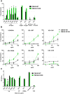
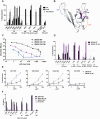
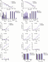
HIV-1 envelope glycoproteins (Env) are the only viral antigens present on the virus surface and serve as the key targets for virus-neutralizing antibodies. However, HIV-1 deploys multiple strategies to shield the vulnerable sites on its Env from neutralizing antibodies. The V1V2 domain located at the apex of the HIV-1 Env spike is known to encompass highly variable loops, but V1V2 also contains immunogenic conserved elements recognized by cross-reactive antibodies. This study evaluates human monoclonal antibodies (mAbs) against V2 epitopes which overlap with the conserved integrin α4β7-binding LDV/I motif, designated as the V2i (integrin) epitopes. We postulate that the V2i Abs have weak or no neutralizing activities because the V2i epitopes are often occluded from antibody recognition. To gain insights into the mechanisms of the V2i occlusion, we evaluated three elements at the distal end of the V1V2 domain shown in the structure of V2i epitope complexed with mAb 830A to be important for antibody recognition of the V2i epitope. Amino-acid substitutions at position 179 that restore the LDV/I motif had minimal effects on virus sensitivity to neutralization by most V2i mAbs. However, a charge change at position 153 in the V1 region significantly increased sensitivity of subtype C virus ZM109 to most V2i mAbs. Separately, a disulfide bond introduced to stabilize the hypervariable region of V2 loop also enhanced virus neutralization by some V2i mAbs, but the effects varied depending on the virus. These data demonstrate that multiple elements within the V1V2 domain act independently and in a virus-dependent fashion to govern the antibody recognition and accessibility of V2i epitopes, suggesting the need for multi-pronged strategies to counter the escape and the shielding mechanisms obstructing the V2i Abs from neutralizing HIV-1.
Conflict of interest statement
Figures


Similar articles
-
Distinct mechanisms regulate exposure of neutralizing epitopes in the V2 and V3 loops of HIV-1 envelope.J Virol. 2014 Nov;88(21):12853-65. doi: 10.1128/JVI.02125-14. Epub 2014 Aug 27. J Virol. 2014. PMID: 25165106 Free PMC article.
-
Plasticity and Epitope Exposure of the HIV-1 Envelope Trimer.J Virol. 2017 Aug 10;91(17):e00410-17. doi: 10.1128/JVI.00410-17. Print 2017 Sep 1. J Virol. 2017. PMID: 28615206 Free PMC article.
-
A Trimeric HIV-1 Envelope gp120 Immunogen Induces Potent and Broad Anti-V1V2 Loop Antibodies against HIV-1 in Rabbits and Rhesus Macaques.J Virol. 2018 Feb 12;92(5):e01796-17. doi: 10.1128/JVI.01796-17. Print 2018 Mar 1. J Virol. 2018. PMID: 29237847 Free PMC article.
-
Vaccine-induced V1V2-specific antibodies control and or protect against infection with HIV, SIV and SHIV.Curr Opin HIV AIDS. 2019 Jul;14(4):309-317. doi: 10.1097/COH.0000000000000551. Curr Opin HIV AIDS. 2019. PMID: 30994501 Free PMC article. Review.
-
[Role of the HIV-1 gp120 V1/V2 domains in the induction of neutralizing antibodies].Enferm Infecc Microbiol Clin. 2009 Nov;27(9):523-30. doi: 10.1016/j.eimc.2008.02.010. Epub 2009 May 1. Enferm Infecc Microbiol Clin. 2009. PMID: 19409660 Review. Spanish.
Cited by
-
P2X1 Selective Antagonists Block HIV-1 Infection through Inhibition of Envelope Conformation-Dependent Fusion.J Virol. 2020 Feb 28;94(6):e01622-19. doi: 10.1128/JVI.01622-19. Print 2020 Feb 28. J Virol. 2020. PMID: 31852781 Free PMC article.
-
Mucosal Delivery of HIV-1 Glycoprotein Vaccine Candidate Enabled by Short Carbon Nanotubes.Part Part Syst Charact. 2022 May;39(5):2200011. doi: 10.1002/ppsc.202200011. Epub 2022 Mar 7. Part Part Syst Charact. 2022. PMID: 36186663 Free PMC article.
-
Single-Cell Transcriptomics of Human Tonsils Reveals Nicotine Enhances HIV-1-Induced NLRP3 Inflammasome and Mitochondrial Activation.Viruses. 2024 Nov 20;16(11):1797. doi: 10.3390/v16111797. Viruses. 2024. PMID: 39599911 Free PMC article.
-
Stabilization of the V2 loop improves the presentation of V2 loop-associated broadly neutralizing antibody epitopes on HIV-1 envelope trimers.J Biol Chem. 2019 Apr 5;294(14):5616-5631. doi: 10.1074/jbc.RA118.005396. Epub 2019 Feb 6. J Biol Chem. 2019. PMID: 30728245 Free PMC article.
-
V2-Specific Antibodies in HIV-1 Vaccine Research and Natural Infection: Controllers or Surrogate Markers.Vaccines (Basel). 2019 Aug 6;7(3):82. doi: 10.3390/vaccines7030082. Vaccines (Basel). 2019. PMID: 31390725 Free PMC article. Review.
References
-
- Karasavvas N, Billings E, Rao M, Williams C, Zolla-Pazner S, Bailer RT, et al. The Thai Phase III HIV Type 1 Vaccine trial (RV144) regimen induces antibodies that target conserved regions within the V2 loop of gp120. AIDS research and human retroviruses. 2012;28(11):1444–57. Epub 2012/10/06. 10.1089/aid.2012.0103 - DOI - PMC - PubMed
Publication types
MeSH terms
Substances
Grants and funding
LinkOut - more resources
Full Text Sources
Other Literature Sources
Medical
Miscellaneous

