. 2015 Oct 22;11(10):e1005203.
doi: 10.1371/journal.ppat.1005203. eCollection 2015 Oct.

Crystal Structures of a Piscine Betanodavirus: Mechanisms of Capsid Assembly and Viral Infection

Affiliations

Crystal Structures of a Piscine Betanodavirus: Mechanisms of Capsid Assembly and Viral Infection

Nai-Chi Chen et al. PLoS Pathog. .

Abstract

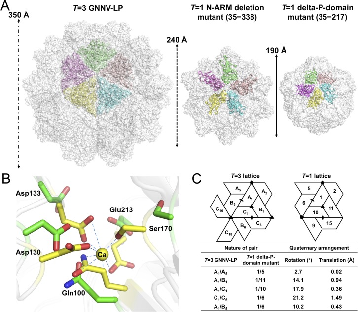
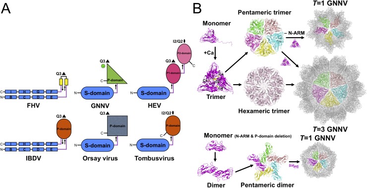
Betanodaviruses cause massive mortality in marine fish species with viral nervous necrosis. The structure of a T = 3 Grouper nervous necrosis virus-like particle (GNNV-LP) is determined by the ab initio method with non-crystallographic symmetry averaging at 3.6 Å resolution. Each capsid protein (CP) shows three major domains: (i) the N-terminal arm, an inter-subunit extension at the inner surface; (ii) the shell domain (S-domain), a jelly-roll structure; and (iii) the protrusion domain (P-domain) formed by three-fold trimeric protrusions. In addition, we have determined structures of the T = 1 subviral particles (SVPs) of (i) the delta-P-domain mutant (residues 35-217) at 3.1 Å resolution; and (ii) the N-ARM deletion mutant (residues 35-338) at 7 Å resolution; and (iii) the structure of the individual P-domain (residues 214-338) at 1.2 Å resolution. The P-domain reveals a novel DxD motif asymmetrically coordinating two Ca2+ ions, and seems to play a prominent role in the calcium-mediated trimerization of the GNNV CPs during the initial capsid assembly process. The flexible N-ARM (N-terminal arginine-rich motif) appears to serve as a molecular switch for T = 1 or T = 3 assembly. Finally, we find that polyethylene glycol, which is incorporated into the P-domain during the crystallization process, enhances GNNV infection. The present structural studies together with the biological assays enhance our understanding of the role of the P-domain of GNNV in the capsid assembly and viral infection by this betanodavirus.


Conflict of interest statement

The authors have declared that no competing interests exist.

Figures

Fig 1
Fig 1. EM image and the overall structure of GNNV-LP.
(A) A representative negative-staining EM image of the purified GNNV-LPs after self-assembly. (B) A ribbon presentation of the subunit C of GNNV-LP. The disordered N-ARM (residue 1−33, gray), N-arm (residues 34−51, magenta), the S-domain (residues 52−213, red), the linker region (residues 214−220, blue), the P-domain (residues 221−338, cyan) and Ca2+ ion (yellow sphere) are shown. (C) A topology diagram of GNNV CP with the helices and strands in cylinders and arrows, respectively. The 1D topology of the subunit C is color-coded as in B. (D) Surface domain-colored diagram (left) and central cavity (right) representations of the T = 3 GNNV-LP. The tip-to-tip distance is ~350 Å, the diameter of the central cavity is ~228 Å, and the spike protrusion on the capsid surface is ~47 Å. The S-domains of the subunits A, B and C are shown in orange, blue and red, respectively, and the P-domains are shown in cyan. The structure of the GNNV-LP is viewed along the I2, I3 and I5 axes.
Fig 2
Fig 2. Structural organizations of the N-arm and the β-annulus.
(A) Two N-arms from the subunit-C/C dimer along the I2 interface. N-arms are colored in magenta. Two iASUs are shown in white and orange. (B) A representation of the hexameric capsomers and the β-annulus as viewed along the I3 axis from the inner surface. Three iASUs are shown in cyan, green and orange, respectively (left). Residues 36−41 of three different C-subunits form a β-annulus structure as an enlarged central section (right). The hydrogen bonds are shown with blue dotted lines. (C) A comparison of the N-arms with an alignment of the S-domains (subunit C) from four different representative viruses: SeMV (green), SMSV (yellow), RyMV (blue) and GNNV (cyan) with a nomenclature diagram. The orientations of the N-arm between GNNV and SeMV are towards opposite different I3 axes (black triangle).
Fig 3
Fig 3. Trimeric interactions of the P-domain with Ca2+ ions and the functional role of PEG during GNNV infection.
(A) The ribbon model of trimeric P-domains. The structure of trimeric P-domains (magenta, green and cyan) is shown with two Ca2+ ions (yellow spheres) bound at the top region. (B) The electrostatic surface potential of the trimeric P-domains. A top view of the trimeric P-domains is rotated 90° along the horizontal axis from A, and colored in red and blue for negative and positive charges viewed along the three-fold axis. (C) The calcium-binding region of the trimeric P-domains. Two Ca2+ ions (yellow spheres) and two water molecules (blue) are coordinated with three sets of 273 DxD 275 motifs (sticks) from neighboring subunits colored in magenta, green and cyan, respectively. The hydrogen bonds are shown with orange dotted lines. (D) Sequence-alignment variables mapped onto the surface of the P-domain from different genotypes of betanodavirus. The hypervariable regions (yellow) from OSGNNV, DGNNV, RGNNV, BFNNV, SJNNV and TPNNV are represented on the surface of the trimeric P-domains (upper). The comparison of representative T4 genomic regions of the P-domains of different genotypes of betanodavirus is shown (lower). Strictly variants and similar residues are colored in yellow (as in D, upper) and green, respectively. (E) Improvement of GNNV infection by PEG treatment. PEG8000 assists GNNV infection in GF-1 cells. Intracellular GNNV RNA2 copies in GF-1 cells were determined by real-time qPCR in log scales after GNNV infection at the indicated time. Data are represented as mean ± SD of three independent experiments and analyzed by one-way ANOVA test, *P < 0.05; **P < 0.01; ***P < 0.001.
Fig 4
Fig 4. T = 1 SVPs of GNNV.
(A) Surface presentations of the monomers engaged in pentameric interactions in the T = 3 and T = 1 GNNV capsids. Each iASU is viewed along the I5 axis and shown in green, wheat, cyan, yellow and magenta colors, respectively. The diameters of three capsids are indicated. (B) A superimposition of the calcium-binding regions of the S-domain between the T = 3 GNNV-LP and the T = 1 delta-P-domain mutant. Gln100, Asp130, Asp133, Ser170 and Glu213, which participate in the Ca2+ coordination in the T = 3 GNNV-LP (yellow), exhibit conformational changes in the T = 1 delta-P-domain mutant (green). The hydrogen bonds are indicated with blue dotted lines. (C) A comparison of the spatial relationship of subunit pairs in the T = 3 GNNV-LP and the T = 1 delta-P-domain mutant. Subunit packing and the nomenclature of the T = 3 and the T = 1 capsids are shown. Rotation angles (°) and translation distances (Å) are identified and compared between different subunit pairs of the T = 3 and the T = 1 GNNV capsids.
Fig 5
Fig 5. Evolutionary lineages and self-assembly mechanisms of GNNV.
(A) Structural characteristics of different categories of RNA viruses. Simplified topology cartoons (β-strand, rectangles; jelly-roll structure, capsules; β-barrel; ovals) of CPs from different viruses, including FHV, IBDV, GNNV, Orsay virus, HEV and tombusvirus are shown. S-domains are colored in blue; P-domains are colored in smudge, gray and orange, respectively; the P1- and P2-domains are indicated in magenta and pink, respectively. The Ca2+ ion in the P-domain of GNNV is labeled as a green sphere. The direction (from N- to C-terminal) of the linker region of each CP is indicated with arrows. (B) Diagram showing the putative self-assembly process of the T = 3 and the T = 1 GNNV capsids. The P-domain and Ca2+ ions (yellow spheres) encode information that controls trimerization of the GNNV CPs for the putative pentameric and hexameric trimers. The N-ARM guides the assembly of the complete T = 3 capsid.

References

    1. Ahlquist P. RNA-dependent RNA polymerases, viruses, and RNA silencing. Science. 2002;296: 1270–1273. - PubMed
    1. Ahlquist P. Parallels among positive-strand RNA viruses, reverse-transcribing viruses and double-stranded RNA viruses. Nat Rev Microbiol. 2005;4: 371–382. - PMC - PubMed
    1. Chao JA, Lee JH, Chapados BR, Debler EW, Schneemann A, Williamson JR. Dual modes of RNA-silencing suppression by Flock house virus protein B2. Nat Struct Mol Biol. 2006;12: 952–957. - PubMed
    1. Li Y, Lu JF, Han YH, Fan XX, Ding SW. RNA interference functions as an antiviral immunity mechanism in mammals. Science. 2013;342: 231–234. 10.1126/science.1241911 - DOI - PMC - PubMed
    1. Lu R, Maduro M, Li F, Li HW, Broitman-Maduro G, Li WX, et al. Animal virus replication and RNAi-mediated antiviral silencing in Caenorhabditis elegans . Nature. 2005;436: 1040–1043. - PMC - PubMed

Publication types

LinkOut - more resources