

Optimized (31)P MRS in the human brain at 7 T with a dedicated RF coil setup
- PMID: 26492089
- PMCID: PMC4744789
- DOI: 10.1002/nbm.3422
Optimized (31)P MRS in the human brain at 7 T with a dedicated RF coil setup
Abstract
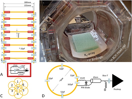
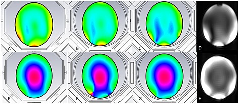
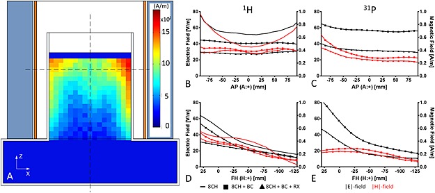
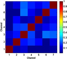
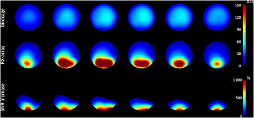
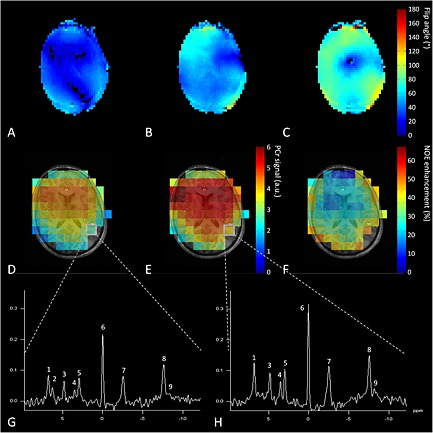
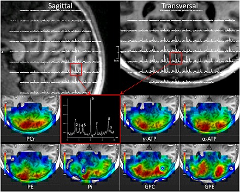
The design and construction of a dedicated RF coil setup for human brain imaging ((1)H) and spectroscopy ((31)P) at ultra-high magnetic field strength (7 T) is presented. The setup is optimized for signal handling at the resonance frequencies for (1)H (297.2 MHz) and (31)P (120.3 MHz). It consists of an eight-channel (1)H transmit-receive head coil with multi-transmit capabilities, and an insertable, actively detunable (31)P birdcage (transmit-receive and transmit only), which can be combined with a seven-channel receive-only (31)P array. The setup enables anatomical imaging and (31)P studies without removal of the coil or the patient. By separating transmit and receive channels and by optimized addition of array signals with whitened singular value decomposition we can obtain a sevenfold increase in SNR of (31)P signals in the occipital lobe of the human brain compared with the birdcage alone. These signals can be further enhanced by 30 ± 9% using the nuclear Overhauser effect by B1-shimmed low-power irradiation of water protons. Together, these features enable acquisition of (31)P MRSI at high spatial resolutions (3.0 cm(3) voxel) in the occipital lobe of the human brain in clinically acceptable scan times (~15 min).
Keywords: 31P-MRS; 31P-MRSI; 7 T; B1 shimming; RF coil; WSVD; array coil; multi-transmit; spectroscopic imaging; ultra-high field.
© 2015 The Authors. NMR in Biomedicine published by John Wiley & Sons Ltd.
Figures
Similar articles
-
Whole-body radiofrequency coil for (31) P MRSI at 7 T.NMR Biomed. 2016 Jun;29(6):709-20. doi: 10.1002/nbm.3517. Epub 2016 Apr 1. NMR Biomed. 2016. PMID: 27037615
-
Eight-channel phased array coil and detunable TEM volume coil for 7 T brain imaging.Magn Reson Med. 2005 Jul;54(1):235-40. doi: 10.1002/mrm.20547. Magn Reson Med. 2005. PMID: 15968650 Clinical Trial.
-
(1) H MRS in the human spinal cord at 7 T using a dielectric waveguide transmitter, RF shimming and a high density receive array.NMR Biomed. 2016 Sep;29(9):1231-9. doi: 10.1002/nbm.3541. Epub 2016 May 18. NMR Biomed. 2016. PMID: 27191947
-
RF pulse methods for use with surface coils: Frequency-modulated pulses and parallel transmission.J Magn Reson. 2018 Jun;291:84-93. doi: 10.1016/j.jmr.2018.01.012. Epub 2018 Apr 26. J Magn Reson. 2018. PMID: 29705035 Free PMC article. Review.
-
Phosphorus-31: A table-top method for 3D B1-field amplitude and phase measurements.Biochim Biophys Acta Biomembr. 2024 Apr;1866(4):184307. doi: 10.1016/j.bbamem.2024.184307. Epub 2024 Feb 24. Biochim Biophys Acta Biomembr. 2024. PMID: 38408694 Review.
Cited by
-
SNR optimized 31 P functional MRS to detect mitochondrial and extracellular pH change during visual stimulation.NMR Biomed. 2019 Nov;32(11):e4137. doi: 10.1002/nbm.4137. Epub 2019 Jul 22. NMR Biomed. 2019. PMID: 31329342 Free PMC article.
-
(31)P CSI of the human brain in healthy subjects and tumor patients at 9.4 T with a three-layered multi-nuclear coil: initial results.MAGMA. 2016 Jun;29(3):579-89. doi: 10.1007/s10334-016-0524-9. Epub 2016 Jan 25. MAGMA. 2016. PMID: 26811174
-
RF Coil Setup for 31P MRSI in Tongue Cancer in vivo at 7 T.Front Neurol. 2021 Nov 2;12:695202. doi: 10.3389/fneur.2021.695202. eCollection 2021. Front Neurol. 2021. PMID: 34795625 Free PMC article.
-
Imaging human teeth by phosphorus magnetic resonance with nuclear Overhauser enhancement.Sci Rep. 2016 Aug 8;6:30756. doi: 10.1038/srep30756. Sci Rep. 2016. PMID: 27498919 Free PMC article.
-
3D 31 P MR spectroscopic imaging of the human brain at 3 T with a 31 P receive array: An assessment of 1 H decoupling, T1 relaxation times, 1 H-31 P nuclear Overhauser effects and NAD.NMR Biomed. 2021 May;34(5):e4169. doi: 10.1002/nbm.4169. Epub 2019 Sep 13. NMR Biomed. 2021. PMID: 31518036 Free PMC article.
References
-
- Hugg JW, Matson GB, Twieg DB, Maudsley AA, Sappey‐Marinier D, Weiner MW. Phosphorus‐31 MR spectroscopic imaging (MRSI) of normal and pathological human brains. Magn. Reson. Imaging 1992; 10(2): 227–243. - PubMed
-
- Wright SM, Wald LL. Theory and application of array coils in MR spectroscopy. NMR Biomed. 1997; 10(8): 394–410. - PubMed
-
- Tannus A, Garwood M. Adiabatic pulses. NMR Biomed. 1997; 10(8): 423–434. - PubMed
Publication types
MeSH terms
Substances
Grants and funding
LinkOut - more resources
Full Text Sources
Other Literature Sources
Medical
