. 2016;48(3):177-88.
doi: 10.3109/23744235.2015.1100323. Epub 2015 Oct 22.

Highly differentiated human airway epithelial cells: a model to study host cell-parasite interactions in pertussis

Affiliations

Highly differentiated human airway epithelial cells: a model to study host cell-parasite interactions in pertussis

Claudia Guevara et al. Infect Dis (Lond). 2016.

Abstract

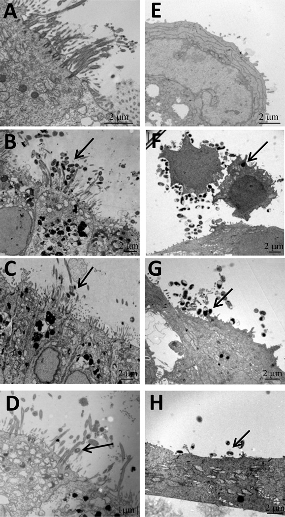
Background: Bordetella pertussis colonizes the human respiratory mucosa. Most studies on B. pertussis adherence have relied on cultured mammalian cells that lack key features present in differentiated human airway cells or on animal models that are not natural hosts of B. pertussis. The objectives of this work were to evaluate B. pertussis infection in highly differentiated human airway cells in vitro and to show the role of B. pertussis fimbriae in cell adherence.

Methods: Primary human airway epithelial (PHAE) cells from human bronchi and a human bronchial epithelial (HBE) cell line were grown in vitro under air-liquid interface conditions.

Results: PHAE and HBE cells infected with B. pertussis wild-type strain revealed bacterial adherence to the apical surface of cells, bacteria-induced cytoskeleton changes, and cell detachment. Mutations in the major fimbrial subunits Fim2/3 or in the minor fimbrial adhesin subunit FimD affected B. pertussis adherence to predominantly HBE cells. This cell model recapitulates the morphologic features of the human airway infected by B. pertussis and confirms the role of fimbriae in B. pertussis adherence. Furthermore, HBE cells show that fimbrial subunits, and specifically FimD adhesin, are critical in B. pertussis adherence to airway cells.

Conclusions: The relevance of this model to study host-parasite interaction in pertussis lies in the striking physiologic and morphologic similarity between the PHAE and HBE cells and the human airway ciliated and goblet cells in vivo. These cells can proliferate in vitro, differentiate, and express the same genetic profile as human respiratory cells in vivo.

Keywords: Bordetella pertussis; adherence; fimbriae major subunit Fim2 or Fim3; minor subunit FimD.


Figures

Figure 1
Figure 1. Primary human airway epithelial (PHAE) cells infected with B. pertussis wild-type strain
PHAE cells were infected with B. pertussis BP536 Tohama I wild-type strain for 6 h before processing for microscopy. Panels A and E, uninfected PHAE cells. Panels B, C, D, and F, cells infected with B. pertussis wild-type strain. Panels A, B, C and D are transmission electron-micrographs. Panels E and F are toluidine blue stained light micrographs. Arrows in panel B, C and D indicate B. pertussis. Arrows in panel F indicate cell detachment and cell flattening.
Figure 2
Figure 2. Human airway epithelial (HBE) cells infected with B. pertussis wild-type strain
HBE cells were infected with B. pertussis BP536 Tohama I wild-type strain for 6 h before processing for microscopy. Panels A and E, are uninfected HBE control cells; panels B, C, D and F are cells infected with B. pertussis. Panels A, B, C and D are transmission electron-micrographs. Panels E and F are haematoxilin/eosin light micrographs. Arrows in panels B, C and D indicate B. pertussis. Arrow in panel F indicate cell detachment after B. pertussis infection.
Figure 3
Figure 3. Bacterial-cell association assays using epithelial cells infected with B. pertussis strains
Panel A. Fimbriae-mediated B. pertussis adherence to epithelial cells. PHAE, HBE, and HeLa cells infected with B. pertussis BP536 Tohama I wild-type strain for 6h were used for quantitative adherence assays. Cell infections were conducted in duplicate. Number of colony forming units per mL (CFU/mL) results are shown (logarithmic scale). WT: B. pertussis BP536 wild-type strain (B. pertussis Tohama I derivative). Fim2/3: fim2/3 B. pertussis mutant; fim2/3-fimD: B. pertussis BP536 fim2/3-fimD mutant. The difference in adherence between wild-type and fim2/3-fimD mutant for each type of PHAE, HBE, and HeLa cells was statistically significant (p<0.05). The difference in adherence between wild type and the fim2/3 mutant was only significant with HBE and HeLa cells (p<0.05), not for PHAE cells. This figure is a representative experiment conducted in duplicate and repeated two to three times. Asterisks on B. pertussis fimbrial mutants represent statistically significant differences in adherence to cells when compared to B. pertussis wild type. Panel B. Immunoblots to evaluate expression of Fim2 and FimD proteins among B. pertussis wild-type strains (Tohama and BP536), B. pertussis fim2/3 mutant strain, or B. pertussis fim2/3-fimD mutant strain. Proteins are derived from whole cell lysates that after separation in an SDS-PAGE, were transferred to a membrane and evaluated with anti-Fim2 or anti-FimD mouse polyclonal antibodies. Arrow indicates the location of the protein of interest and the corresponding mass.
Figure 4
Figure 4. Fim2 and FimD proteins surface exposure by B. pertussis
Confocal Laser Scanning Microscopical (CLSM) Analyses were conducted to evaluate the presence of Fim2 or FimD proteins on the surface of B. pertussis strains. Mouse anti-Fim2 polyclonal antibody or mouse anti-FimD polyclonal antibodies were used to label negative control E. coli DH5α, B. pertussis wild-type strain, B. pertussis fim2/3 mutant strain, or B. pertussis fim2/3-fimD mutant strain. Images were taken at 630× magnification and scale bars indicate 50 µm.
Figure 5
Figure 5. PHAE and HBE cells infected with B. pertussis wild-type and fimbrial mutant strains
HBE cells were infected for 6 h with B. pertussis wild-type strain, fim2/3 mutant, or fim2/3-fimD mutant strains before processing for transmission electron-microscopy as described in material and methods. Panels A to D show PHAE cells; panels E to H, HBE cells; panel A and E, uninfected cells; panels B and F, B. pertussis wild-type; panel C and G, B. pertussis fim2/3 mutant strain; panel D and H, B. pertussis fim2/3-fimD mutant strain.
Figure 6
Figure 6. Bacterial-cell association assay using HBE cells infected with B. pertussis strains in the presence of fimbrial inhibitors
HBE cells were infected with B. pertussis wild-type strain, fim2/3 mutant or fim2/3-fimD mutant in the presence of increasing concentration of purified B. pertussis fimbriae proteins Fim2/3 and FimD and enterotoxigenic E. coli CS21 fimbriae LngA protein, as control. Cell infections at each inhibitor concentration were conducted in duplicate. Difference in adherence to cells between wild type and fimbrial mutants was statistically significant (p<0.01) only in the absence or, in some cases, at low concentrations of Fim2/3 and FimD inhibitors. Differences in adherence between wild type and fimbrial mutant were always statistically significant (p<0.01) regardless the presence or absence of LngA inhibitor. This figure show representative experiments conducted in duplicate and repeated two to three times. Asterisk represents statistically significant differences in wild type adherence to cells with respect to fimbrial mutants.

Similar articles

Cited by

References

    1. Melvin JA, Scheller EV, Miller JF, Cotter PA. Bordetella pertussis pathogenesis: current and future challenges. Nat Rev Microbiol. 2014;12(4):274–288. - PMC - PubMed
    1. Mattoo S, Cherry JD. Molecular pathogenesis, epidemiology, and clinical manifestations of respiratory infections due to Bordetella pertussis and other Bordetella subspecies. Clin Microbiol Rev. 2005;18(2):326–382. - PMC - PubMed
    1. Surridge J, Segedin ER, Grant CC. Pertussis requiring intensive care. Arch Dis Child. 2007;92(11):970–975. - PMC - PubMed
    1. Burns DL, Meade BD, Messionnier NE. Pertussis resurgence: perspectives from the Working Group Meeting on pertussis on the causes, possible paths forward, and gaps in our knowledge. J Infect Dis. 2014;209(Suppl 1):S32–S35. - PubMed
    1. Crowcroft NS, Stein C, Duclos P, Birmingham M. How best to estimate the global burden of pertussis? Lancet Infect Dis. 2003;3(7):413–418. - PubMed

Publication types