. 2015 Oct 22:5:15022.
doi: 10.1038/srep15022.

Sickle cell detection using a smartphone

Affiliations

Sickle cell detection using a smartphone

S M Knowlton et al. Sci Rep. .

Abstract

Sickle cell disease affects 25% of people living in Central and West Africa and, if left undiagnosed, can cause life threatening "silent" strokes and lifelong damage. However, ubiquitous testing procedures have yet to be implemented in these areas, necessitating a simple, rapid, and accurate testing platform to diagnose sickle cell disease. Here, we present a label-free, sensitive, and specific testing platform using only a small blood sample (<1 μl) based on the higher density of sickle red blood cells under deoxygenated conditions. Testing is performed with a lightweight and compact 3D-printed attachment installed on a commercial smartphone. This attachment includes an LED to illuminate the sample, an optical lens to magnify the image, and two permanent magnets for magnetic levitation of red blood cells. The sample is suspended in a paramagnetic medium with sodium metabisulfite and loaded in a microcapillary tube that is inserted between the magnets. Red blood cells are levitated in the magnetic field based on equilibrium between the magnetic and buoyancy forces acting on the cells. Using this approach, we were able to distinguish between the levitation patterns of sickle versus control red blood cells based on their degree of confinement.


Conflict of interest statement

A provisional patent has been filed on the technology described here (#62133031).

Figures
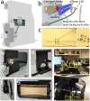
Figure 1
Figure 1. Magnetic levitation based platform (Sickle Cell Tester) installed on a smartphone and optical microscope for static separation of cells.
(a) A schematic diagram of the lightweight smartphone attachment of the Sickle Cell Tester is illustrated. The smartphone attachment is composed of a 3D-printed holder, optical components (aspheric lens and holder), LED illumination, and two permanent magnets with the same poles facing each other. A capillary may be inserted between the magnets by the user. (b) The sample is illuminated with an LED through a ground glass diffuser. The image is magnified by the aspheric lens before it is captured by the built-in smartphone camera (not pictured). (c) Captured image of magnetic levitation of 10 μm polystyrene microspheres (scale bar is 25 μm). (df) Front, back, and side view images of the Sickle Cell Tester platform. (g) An image of levitating microspheres captured with the smartphone. (h) A light microscope laid on its side that allows imaging of levitating cells using an comparable two-magnet configuration to the one shown in (a).
Figure 2
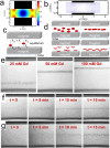
Figure 2. Theoretical and experimental magnetic levitation of RBCs.
(a) Contour plot (b) magnetic field lines representing the magnetic field between the two magnets with like poles facing each other. The magnitude of the magnetic field is constrained between 0 T and 0.4 T. (c) Until the object reaches equilibrium, fluidic drag (Fd), inertial (Fi), buoyancy (Fg), and magnetic forces (Fm) act on the object. As the object gets closer to the equilibrium between Fg and Fm, its migration velocity, and thus drag and inertial forces, become smaller. (d) Theoretical levitation of control RBCs (top) and SS RBCs (bottom), where some SS RBCs levitate at a lower height than control RBCs due to their higher density. (e) Levitation of RBCs in different concentrations of gadolinium solution, demonstrating the effect of the relative magnetic susceptibility on RBC levitation. Time-dependent confinement of (f) control and (g) SS RBCs toward equilibrium in a 50 mM Gd solution with 10 mM sodium metabisulfite.
Figure 3
Figure 3. Cell levitation analysis application running on an Android phone is used to demonstrate the effect of gadolinium concentration on levitation height.
(ad) Images of levitating microspheres are acquired by the user and rapidly processed by the application to determine the levitation height (‘mean’) and confinement width (‘std’) of the microspheres in the sample. (a) 25 mM, (b) 50 mM, (c) 100 mM, and (d) 200 mM gadolinium solutions are tested. (e) The levitation height results (‘mean’, blue) show the varying levitation of microspheres in different concentrations of gadolinium (values are measured relative to the top magnet). The Gaussian distribution results (‘std’, gray) show that the width of confinement is fairly constant across all concentrations of gadolinium with a slight decrease in the width of confinement and a decrease in the variation between samples at higher concentrations of gadolinium. Error bars represent the standard deviation over 6 trials.
Figure 4
Figure 4. Identification of sickle cell disease using the magnetic levitation platform.
Levitation of (a) control and (b) SS RBCs in 50 mM gadolinium solution with 10 mM sodium metabisulfite using a microscopy compatible-setup (Fig. 1h). Levitation of (c) control and (d) SS RBCs in 50 mM gadolinium solution with 10 mM sodium metabisulfite using the Sickle Cell Tester installed on a smartphone. (e) Distribution of Wilcoxon scores for confinement width of 4 control and 11 SS RBC samples analyzed using the Sickle Cell Tester. Results show a statistically significant difference between the groups according to a two-tailed non-parametric Mann-Whitney-Wilcoxon test with a t-approximation (α = 0.05). The full distribution of confinement widths are given in Supplementary Figure 2.

References

    1. World Health Organization. Fifty-Ninth World Health Assembly Resolutions and Decisions Annexes. < http://apps.who.int/gb/ebwha/pdf_files/WHA59-REC1/e/WHA59_2006_REC1-en.pdf> (2006) Access date: 06/30/2015.
    1. Sickle Cell Disease Association of America. Sickle Cell Disease Global: Sickle Cell Disease is a Global Public Health Issue, < http://www.sicklecelldisease.org/index.cfm?page=scd-global> (2014) Access date: 06/30/2015.
    1. National Heart Lung and Blood Institute. What is Sickle Cell Anemia?, < http://www.nhlbi.nih.gov/health/health-topics/topics/sca/#> (2012) Access date: 06/30/2015.
    1. Finch J. T., Perutz M. F., Bertles J. F. & Döbler J. Structure of Sickled Erythrocytes and of Sickle-Cell Hemoglobin Fibers. Proceedings of the National Academy of Sciences 70, 718–722, 10.1073/pnas.70.3.718 (1973). - DOI - PMC - PubMed
    1. Center for Disease Control and Prevention. Facts About Sickle Cell Disease, < http://www.cdc.gov/ncbddd/sicklecell/facts.html> (2014) Access date: 06/30/2015.

Publication types