

Rationale, Design, and Methodological Aspects of the BUDAPEST-GLOBAL Study (Burden of Atherosclerotic Plaques Study in Twins-Genetic Loci and the Burden of Atherosclerotic Lesions)
- PMID: 26492817
- PMCID: PMC6490834
- DOI: 10.1002/clc.22482
Rationale, Design, and Methodological Aspects of the BUDAPEST-GLOBAL Study (Burden of Atherosclerotic Plaques Study in Twins-Genetic Loci and the Burden of Atherosclerotic Lesions)
Abstract
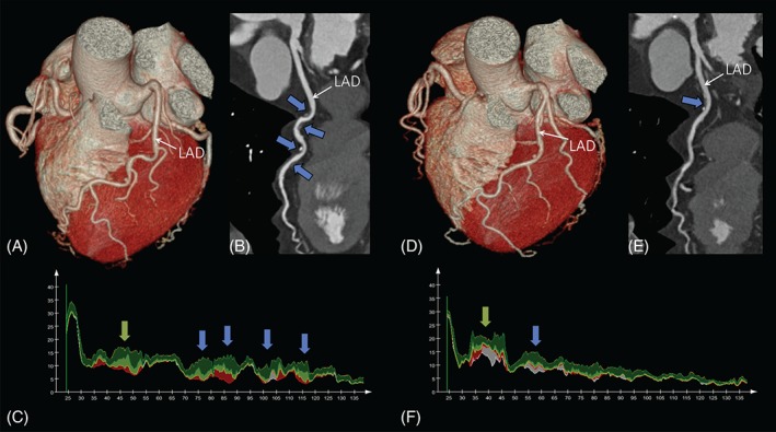
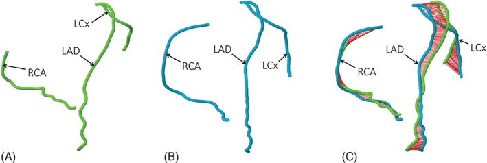
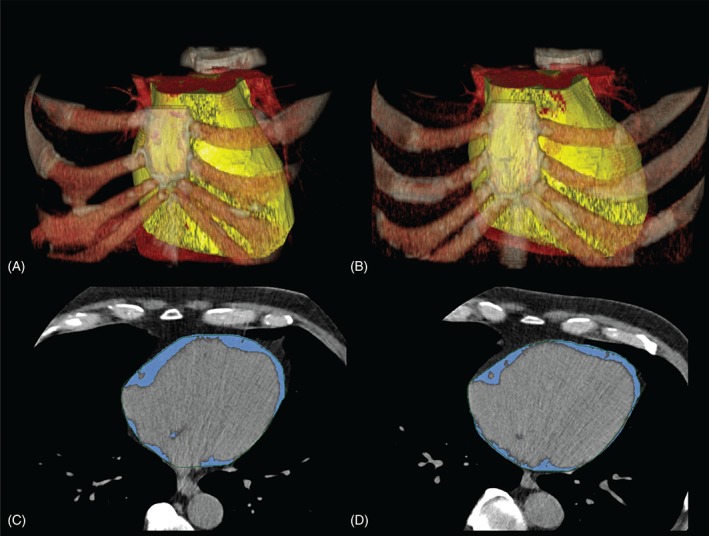
The heritability of coronary atherosclerotic plaque burden, coronary geometry, and phenotypes associated with increased cardiometabolic risk are largely unknown. The primary aim of the Burden of Atherosclerotic Plaques Study in Twins-Genetic Loci and the Burden of Atherosclerotic Lesions (BUDAPEST-GLOBAL) study is to evaluate the influence of genetic and environmental factors on the burden of coronary artery disease. By design this is a prospective, single-center, classical twin study. In total, 202 twins (61 monozygotic pairs, 40 dizygotic same-sex pairs) were enrolled from the Hungarian Twin Registry database. All twins underwent non-contrast-enhanced computed tomography (CT) for the detection and quantification of coronary artery calcium and for the measurement of epicardial fat volumes. In addition, a single non-contrast-enhanced image slice was acquired at the level of L3-L4 to assess abdominal fat distribution. Coronary CT angiography was used for the detection and quantification of plaque, stenosis, and overall coronary artery disease burden. For the primary analysis, we will assess the presence and volume of atherosclerotic plaques. Furthermore, the 3-dimensional coronary geometry will be assessed based on the coronary CT angiography datasets. Additional phenotypic analyses will include per-patient epicardial and abdominal fat quantity measurements. Measurements obtained from monozygotic and dizygotic twin pairs will be compared to evaluate the genetic or environmental effects of the given phenotype. The BUDAPEST-GLOBAL study provides a unique framework to shed some light on the genetic and environmental influences of cardiometabolic disorders.
© 2015 Wiley Periodicals, Inc.
Figures
Similar articles
-
Heritability of Coronary Artery Disease: Insights From a Classical Twin Study.Circ Cardiovasc Imaging. 2022 Mar;15(3):e013348. doi: 10.1161/CIRCIMAGING.121.013348. Epub 2022 Mar 15. Circ Cardiovasc Imaging. 2022. PMID: 35290075 Free PMC article.
-
Precision phenotyping, panomics, and system-level bioinformatics to delineate complex biologies of atherosclerosis: rationale and design of the "Genetic Loci and the Burden of Atherosclerotic Lesions" study.J Cardiovasc Comput Tomogr. 2014 Nov-Dec;8(6):442-51. doi: 10.1016/j.jcct.2014.08.006. Epub 2014 Sep 6. J Cardiovasc Comput Tomogr. 2014. PMID: 25439791
-
Comparison of Quantity of Coronary Atherosclerotic Plaques Detected by Computed Tomography Versus Angiography.Am J Cardiol. 2016 Jun 15;117(12):1863-7. doi: 10.1016/j.amjcard.2016.03.031. Epub 2016 Apr 5. Am J Cardiol. 2016. PMID: 27134058 Clinical Trial.
-
Plaque assessment by coronary CT.Int J Cardiovasc Imaging. 2016 Jan;32(1):161-72. doi: 10.1007/s10554-015-0741-8. Epub 2015 Aug 18. Int J Cardiovasc Imaging. 2016. PMID: 26280890 Review.
-
Genetic and environmental etiology of the relationship between childhood hyperactivity/inattention and conduct problems in a South Korean twin sample.Twin Res Hum Genet. 2015 Jun;18(3):290-7. doi: 10.1017/thg.2015.26. Epub 2015 Apr 30. Twin Res Hum Genet. 2015. PMID: 25926162 Review.
Cited by
-
Left Ventricular Systolic Function Has Strong Independent Genetic Background from Diastolic Function: A Classical Twin Study.Medicina (Kaunas). 2021 Sep 5;57(9):935. doi: 10.3390/medicina57090935. Medicina (Kaunas). 2021. PMID: 34577858 Free PMC article.
-
State of the art paper: Cardiac computed tomography of the left atrium in atrial fibrillation.J Cardiovasc Comput Tomogr. 2023 May-Jun;17(3):166-176. doi: 10.1016/j.jcct.2023.03.002. Epub 2023 Mar 23. J Cardiovasc Comput Tomogr. 2023. PMID: 36966040 Free PMC article. Review.
-
Standardised computed tomographic assessment of left atrial morphology and tissue thickness in humans.Int J Cardiol Heart Vasc. 2020 Dec 24;32:100694. doi: 10.1016/j.ijcha.2020.100694. eCollection 2021 Feb. Int J Cardiol Heart Vasc. 2020. PMID: 33392384 Free PMC article.
-
Clinical importance of epicardial adipose tissue.Arch Med Sci. 2017 Jun;13(4):864-874. doi: 10.5114/aoms.2016.63259. Epub 2016 Oct 26. Arch Med Sci. 2017. PMID: 28721155 Free PMC article.
-
Aortic root dimensions are predominantly determined by genetic factors: a classical twin study.Eur Radiol. 2017 Jun;27(6):2419-2425. doi: 10.1007/s00330-016-4590-1. Epub 2016 Sep 22. Eur Radiol. 2017. PMID: 27659700
References
-
- Mangino M, Spector T. Understanding coronary artery disease using twin studies. Heart. 2013;99:373–375. - PubMed
-
- Derks EM, Dolan CV, Boomsma DI. A test of the equal environment assumption (EEA) in multivariate twin studies. Twin Res Hum Genet. 2006;9:403–411. - PubMed
-
- Kendler KS, Neale MC, Kessler RC, et al. A test of the equal‐environment assumption in twin studies of psychiatric illness. Behav Genet. 1993;23:21–27. - PubMed
Publication types
MeSH terms
LinkOut - more resources
Full Text Sources
Other Literature Sources
Medical
