

Making maxillary barbels with a proximal-distal gradient of Wnt signals in matrix-bound mesenchymal cells
- PMID: 26492827
- PMCID: PMC4620582
- DOI: 10.1111/ede.12167
Making maxillary barbels with a proximal-distal gradient of Wnt signals in matrix-bound mesenchymal cells
Abstract
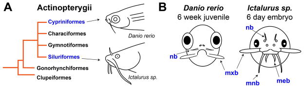
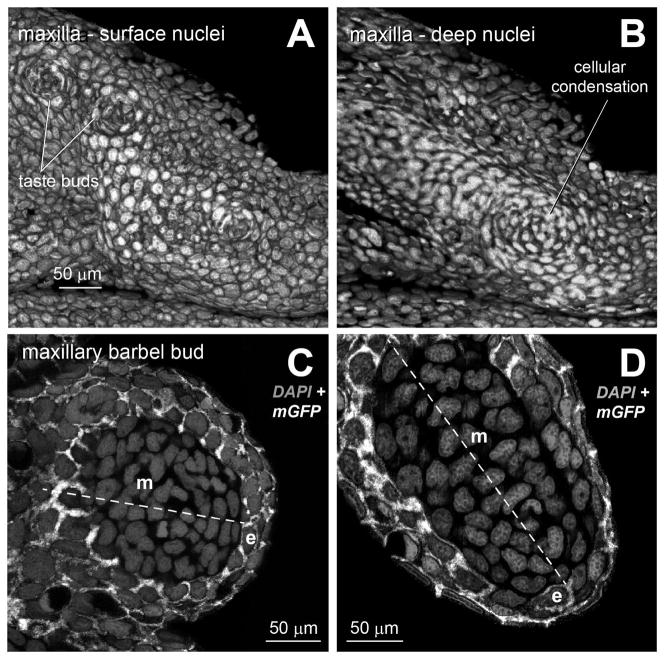
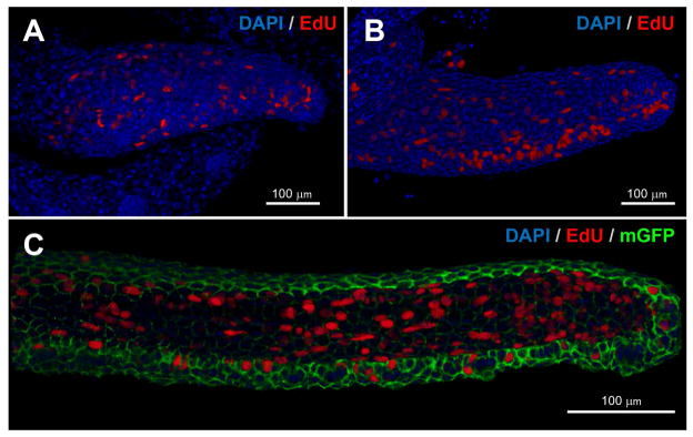
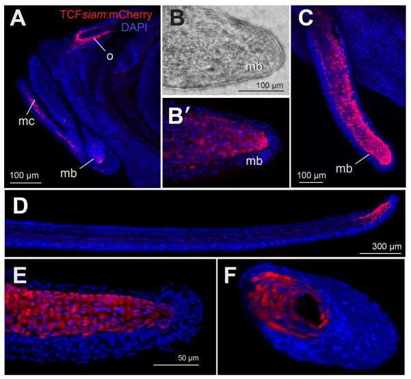
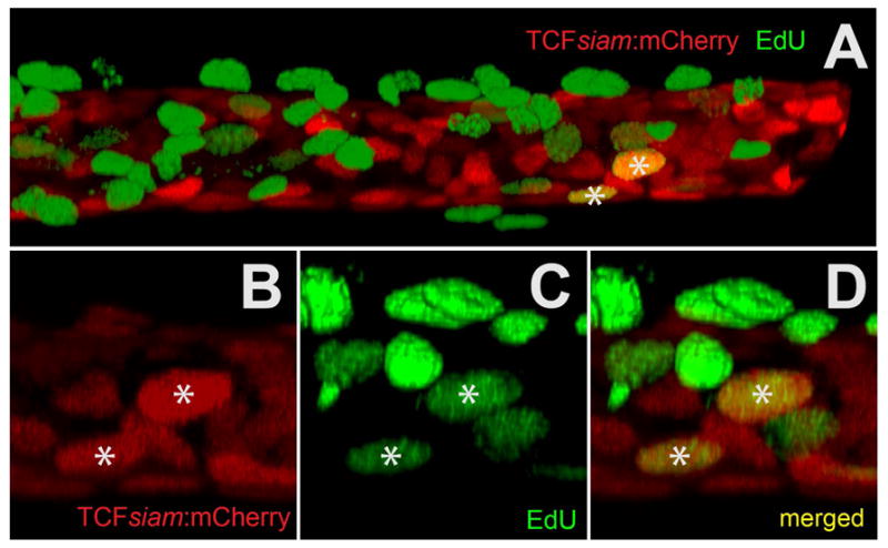
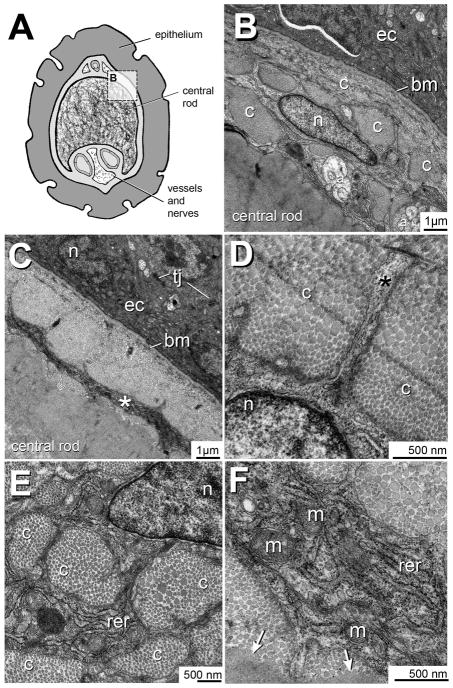
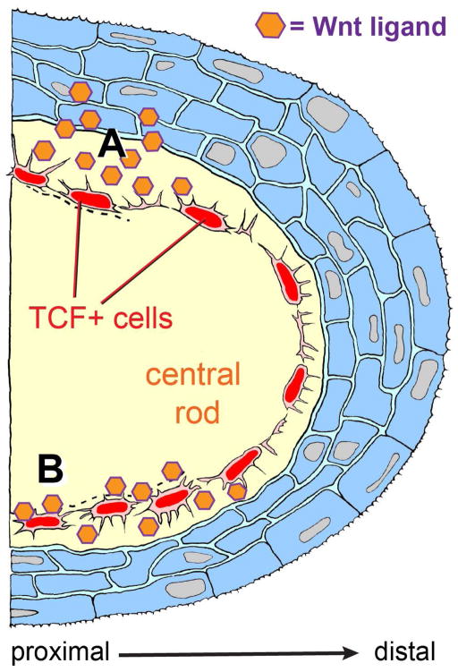
The evolution of specific appendages is made possible by the ontogenetic deployment of general cell signaling pathways. Many fishes, amphibians and reptiles have unique skin appendages known as barbels, which are poorly understood at the cellular and molecular level. In this study, we examine the cell arrangements, cell division patterns, and gene expression profiles associated with the zebrafish maxillary barbel, or ZMB. The earliest cellular organization of the ZMB is an internal whorl of mesenchymal cells in the dermis of the maxilla; there is no epithelial placode, nor any axially-elongated epithelial cells as expected of an apical ectodermal ridge (AER). As the ZMB develops, cells in S-phase are at first distributed randomly throughout the appendage, gradually transitioning to a proliferative population concentrated at the distal end. By observing ZMB ontogenetic stages in a Wnt-responsive transgenic reporter line, TCFsiam, we identified a strongly fluorescent mesenchymal cell layer within these developing appendages. Using an in vitro explant culture technique on developing barbel tissues, we co-localized the fluorescent label in these cells with the mitotic marker EdU. Surprisingly, the labeled cells showed little proliferation, indicating a slow-cycling subpopulation. Transmission electron microscopy of the ZMB located these cells in a single, circumferential layer within the barbel's matrix core. Morphologically, these cells resemble fibroblasts or osteoblasts; in addition to their matrix-bound location, they are identified by their pancake-shaped nuclei, abundant rough endoplasmic reticulum, and cytoplasmic extensions into the surrounding extracellular matrix. Taken together, these features define a novel mesenchymal cell population in zebrafish, the "TCF(+) core cells." A working model of barbel development is proposed, in which these minimally mitotic mesodermal cells produce collagenous matrix in response to ectodermally-derived Wnt signals deployed in a proximal-distal gradient along the appendage. This documents a novel mechanism of vertebrate appendage outgrowth. Similar genetic signals and cell behaviors may be responsible for the independent and repeated evolution of barbel structures in other fish species.
© 2015 Wiley Periodicals, Inc.
Figures
Similar articles
-
Development and regeneration of the zebrafish maxillary barbel: a novel study system for vertebrate tissue growth and repair.PLoS One. 2010 Jan 15;5(1):e8737. doi: 10.1371/journal.pone.0008737. PLoS One. 2010. PMID: 20090899 Free PMC article.
-
Methods for the study of the zebrafish maxillary barbel.J Vis Exp. 2009 Nov 23;(33):1558. doi: 10.3791/1558. J Vis Exp. 2009. PMID: 19935639 Free PMC article.
-
Divergent requirements for fibroblast growth factor signaling in zebrafish maxillary barbel and caudal fin regeneration.Dev Growth Differ. 2013 Feb;55(2):282-300. doi: 10.1111/dgd.12035. Epub 2013 Jan 28. Dev Growth Differ. 2013. PMID: 23350700 Free PMC article.
-
Wnt/planar cell polarity signaling: an important mechanism to coordinate growth and patterning in the limb.Organogenesis. 2011 Oct-Dec;7(4):260-6. doi: 10.4161/org.7.4.19049. Organogenesis. 2011. PMID: 22198433 Free PMC article. Review.
-
Development and evolution of the mammalian limb: adaptive diversification of nails, hooves, and claws.Evol Dev. 2001 Sep-Oct;3(5):355-63. doi: 10.1046/j.1525-142x.2001.01032.x. Evol Dev. 2001. PMID: 11710767 Review.
Cited by
-
Identification of regulatory elements recapitulating early expression of L-plastin in the zebrafish enveloping layer and embryonic periderm.Gene Expr Patterns. 2019 Jun;32:53-66. doi: 10.1016/j.gep.2019.03.001. Epub 2019 Mar 30. Gene Expr Patterns. 2019. PMID: 30940554 Free PMC article.
References
-
- Alibardi L. Dermo-epidermal interactions in reptilian scales: Speculations on the evolution of scales, feathers, and hairs. J Exp Zool B Mol Dev Evol. 2004;302 (4):365–83. - PubMed
-
- Alibardi L. Proliferation in the epidermis of chelonians and growth of the horny scutes. J Morphol. 2005;265 (1):52–69. - PubMed
-
- Ansell DM, Holden KA, Hardman MJ. Animal models of wound repair: Are they cutting it? Exp Dermatol. 2012;21 (8):581–5. - PubMed
-
- Avella M, Berhaut J, Payan P. Primary culture of gill epithelial cells from the sea bass Dicentrarchus labrax. In Vitro Cell Dev Biol Anim. 1994;30A (1):41–9. - PubMed
-
- Baena-López LA, Baonza A, García-Bellido A. The orientation of cell divisions determines the shape of Drosophila organs. Curr Biol. 2005;15 (18):1640–4. - PubMed
Publication types
MeSH terms
Grants and funding
LinkOut - more resources
Full Text Sources
Other Literature Sources
Molecular Biology Databases
Research Materials
