

Non-invasive PET Imaging of PARP1 Expression in Glioblastoma Models
- PMID: 26493053
- PMCID: PMC4841747
- DOI: 10.1007/s11307-015-0904-y
Non-invasive PET Imaging of PARP1 Expression in Glioblastoma Models
Abstract
Purpose: The current study presents [(18)F]PARPi as imaging agent for PARP1 expression.
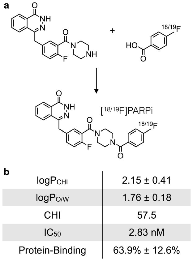
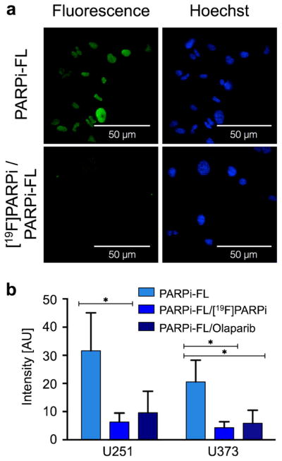
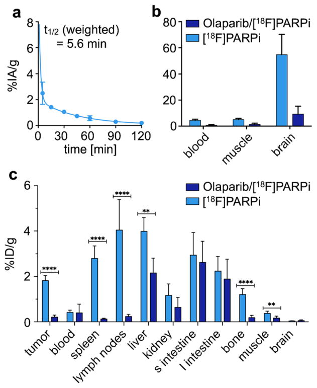
Procedures: [(18)F]PARPi was generated by conjugating a 2H-phthalazin-1-one scaffold to 4-[(18)F]fluorobenzoic acid. Biochemical assays, optical in vivo competition, biodistribution analysis, positron emission tomography (PET)/X-ray computed tomography, and PET/magnetic resonance imaging studies were performed in subcutaneous and orthotopic mouse models of glioblastoma.
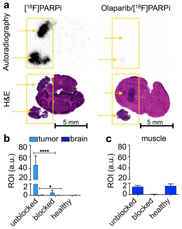
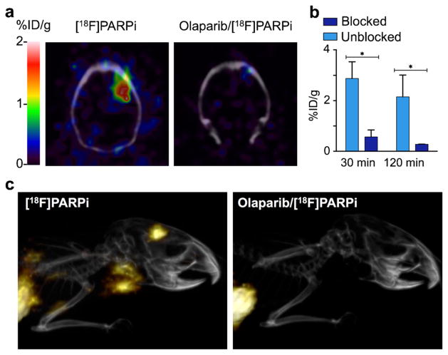
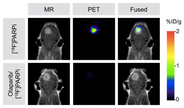
Results: [(18)F]PARPi shows suitable pharmacokinetic properties for brain tumor imaging (IC50 = 2.8 ± 1.1 nM; logPCHI = 2.15 ± 0.41; plasma-free fraction = 63.9 ± 12.6 %) and accumulates selectively in orthotopic brain tumor tissue. Tracer accumulation in subcutaneous brain tumors was 1.82 ± 0.21 %ID/g, whereas in healthy brain, the uptake was only 0.04 ± 0.01 %ID/g.
Conclusions: [(18)F]PARPi is a selective PARP1 imaging agent that can be used to visualize glioblastoma in xenograft and orthotopic mouse models with high precision and good signal/noise ratios. It offers new opportunities to non-invasively image tumor growth and monitor interventions.
Keywords: Glioblastoma; Imaging; Orthotopic; PARP1; PET.
Conflict of interest statement
Figures
Similar articles
-
Dual-Modality Optical/PET Imaging of PARP1 in Glioblastoma.Mol Imaging Biol. 2015 Dec;17(6):848-55. doi: 10.1007/s11307-015-0858-0. Mol Imaging Biol. 2015. PMID: 25895168 Free PMC article.
-
Radioiodinated PARP1 tracers for glioblastoma imaging.EJNMMI Res. 2015 Dec;5(1):123. doi: 10.1186/s13550-015-0123-1. Epub 2015 Sep 4. EJNMMI Res. 2015. PMID: 26337803 Free PMC article.
-
An 18F-Labeled Poly(ADP-ribose) Polymerase Positron Emission Tomography Imaging Agent.J Med Chem. 2018 May 10;61(9):4103-4114. doi: 10.1021/acs.jmedchem.8b00138. Epub 2018 Apr 19. J Med Chem. 2018. PMID: 29630818 Free PMC article.
-
Approaches to PET Imaging of Glioblastoma.Molecules. 2020 Jan 28;25(3):568. doi: 10.3390/molecules25030568. Molecules. 2020. PMID: 32012954 Free PMC article. Review.
-
The Development of 18F Fluorthanatrace: A PET Radiotracer for Imaging Poly (ADP-Ribose) Polymerase-1.Radiol Imaging Cancer. 2022 Jan;4(1):e210070. doi: 10.1148/rycan.210070. Radiol Imaging Cancer. 2022. PMID: 35089089 Free PMC article. Review.
Cited by
-
Biomarker-Based PET Imaging of Diffuse Intrinsic Pontine Glioma in Mouse Models.Cancer Res. 2017 Apr 15;77(8):2112-2123. doi: 10.1158/0008-5472.CAN-16-2850. Epub 2017 Jan 20. Cancer Res. 2017. PMID: 28108511 Free PMC article.
-
Molecular Targets for Pharmacotherapy of Head and Neck Squamous Cell Carcinomas.Curr Issues Mol Biol. 2025 Aug 1;47(8):609. doi: 10.3390/cimb47080609. Curr Issues Mol Biol. 2025. PMID: 40864763 Free PMC article. Review.
-
Perspective on the Use of DNA Repair Inhibitors as a Tool for Imaging and Radionuclide Therapy of Glioblastoma.Cancers (Basel). 2022 Apr 3;14(7):1821. doi: 10.3390/cancers14071821. Cancers (Basel). 2022. PMID: 35406593 Free PMC article. Review.
-
PET imaging of PARP expression using 68Ga-labelled inhibitors.Eur J Nucl Med Mol Imaging. 2023 Jul;50(9):2606-2620. doi: 10.1007/s00259-023-06249-6. Epub 2023 May 5. Eur J Nucl Med Mol Imaging. 2023. PMID: 37145164 Free PMC article.
-
Two experts and a newbie: [18F]PARPi vs [18F]FTT vs [18F]FPyPARP-a comparison of PARP imaging agents.Eur J Nucl Med Mol Imaging. 2022 Feb;49(3):834-846. doi: 10.1007/s00259-021-05436-7. Epub 2021 Sep 6. Eur J Nucl Med Mol Imaging. 2022. PMID: 34486071 Free PMC article.
References
-
- Hassa PO, Hottiger MO. The diverse biological roles of mammalian PARPS, a small but powerful family of poly-ADP-ribose polymerases. Front Biosci. 2008;13:3046–3082. - PubMed
Publication types
MeSH terms
Substances
Grants and funding
LinkOut - more resources
Full Text Sources
Other Literature Sources
Medical
Miscellaneous
