

Evidence from the very beginning: endoglandular trophoblasts penetrate and replace uterine glands in situ and in vitro
- PMID: 26493408
- PMCID: PMC4719185
- DOI: 10.1093/humrep/dev266
Evidence from the very beginning: endoglandular trophoblasts penetrate and replace uterine glands in situ and in vitro
Abstract
Study question: How is histiotrophic nutrition of the embryo secured during the first trimester of pregnancy?
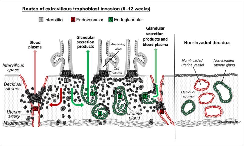
Summary answer: Rather than specifically focusing on invasion into spiral arteries, extravillous trophoblasts also invade into uterine glands (endoglandular trophoblast) from the very beginning and open them toward the intervillous space.
What is known already: Extravillous trophoblasts can be found in close contact and within the lumen of uterine glands, sometimes replacing glandular epithelial cells.
Study design, size, duration: As well as extensive screening of specimens from first trimester placentation sites in situ we used a previously established three-dimensional co-culture in vitro model system of first trimester villous explants with non-invaded decidua parietalis.
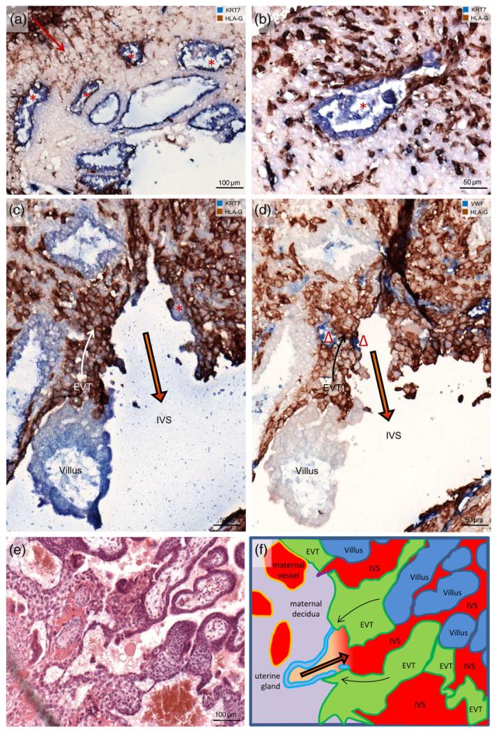
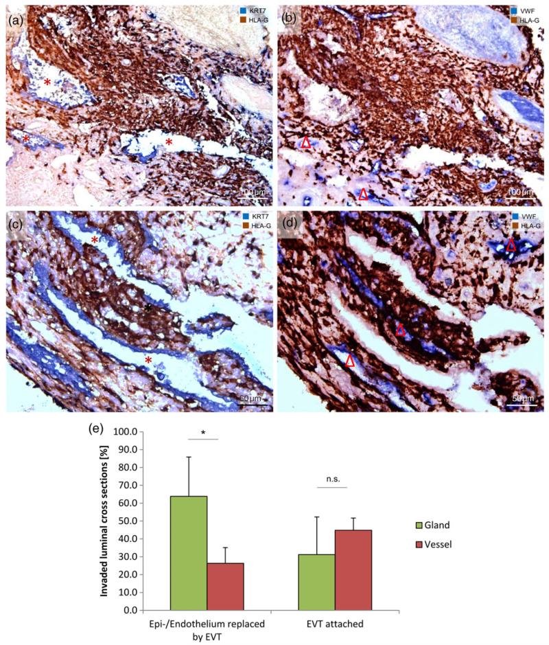
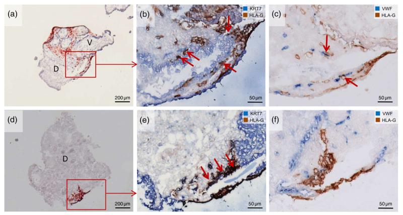
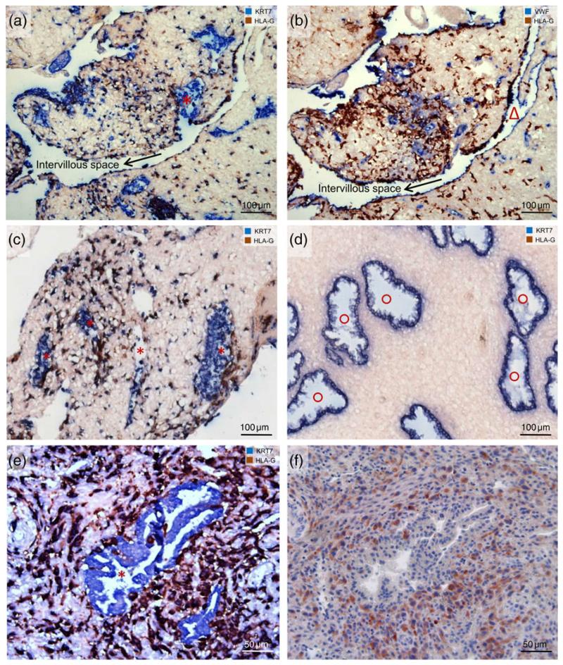
Participants/materials, setting, methods: First trimester placentas were obtained from elective terminations of pregnancies (n = 48) at 5-11 weeks of gestational age. A subset was processed for confrontation co-culture (n = 31). Invaded decidua basalis was obtained from 20 placentas. All tissues were sectioned, subsequently immunostained and immunodoublestained with antibodies against keratin 7 (KRT7), major histocompatibility complex, class I, G (HLA-G), matrix metallopeptidase 9 (MMP9), von Willebrand factor (VWF) and the appropriate Immunoglobulin G (IgG) negative controls. Replacement of endothelial/epithelial cells by extravillous trophoblasts was quantified semi-quantitatively. Additionally, hematoxylin and eosin-stained archival specimens from early implantation sites were assessed.
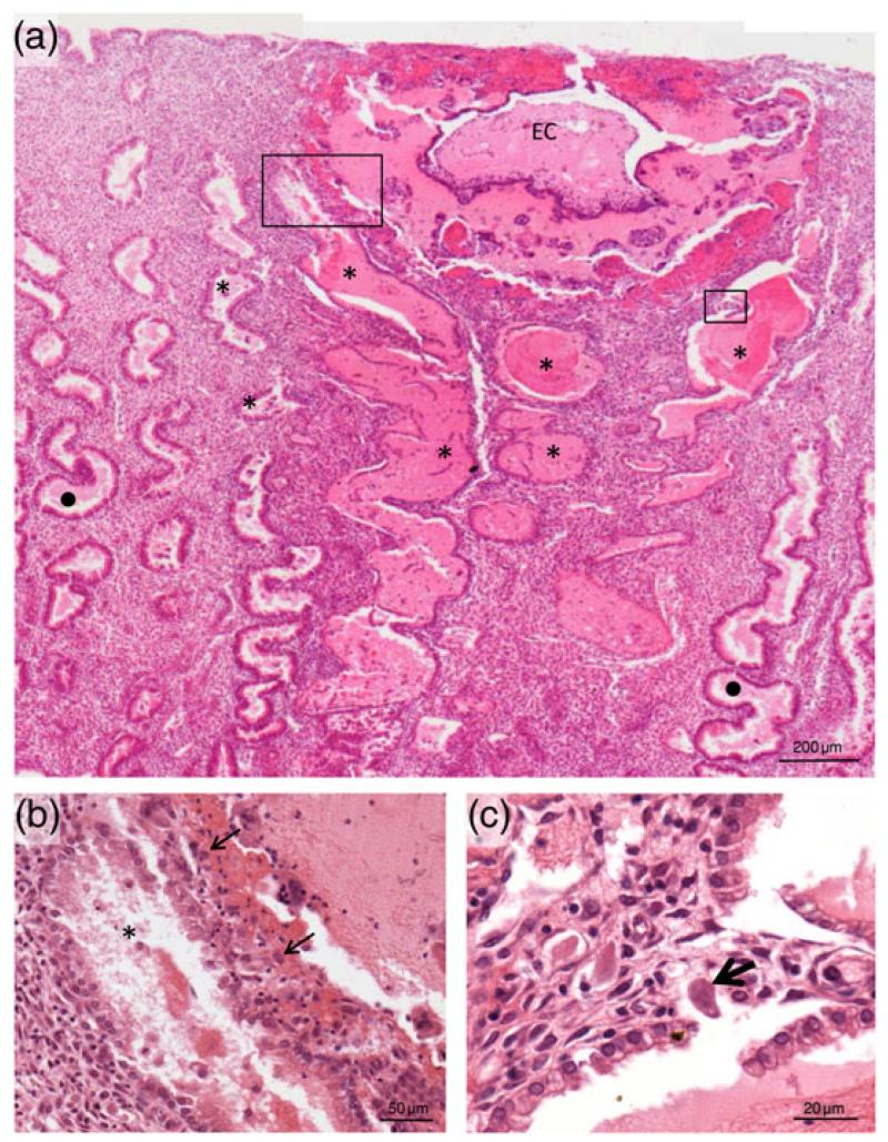
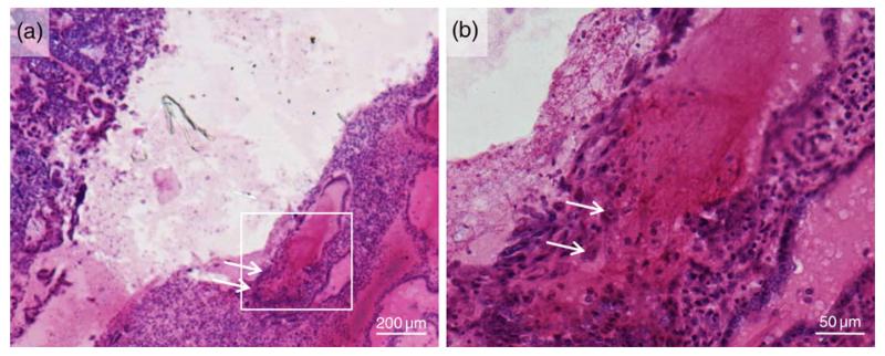
Main results and the role of chance: The earliest available specimen was from around Day 10 after conception; already at this stage trophoblasts had penetrated into uterine glands and had started to replace the epithelium of the glands. Endoglandular trophoblasts replaced uterine glands in vitro and in situ and could be found in the lumen of invaded glands. Quantitative analysis revealed significantly more replacement of epithelial cells in glands (63.8 ± 22.1%) compared with endothelial cells in vessels (26.4 ± 8.8%). Accumulated detached glandular epithelial cells could be repeatedly observed in the lumen of invaded glands. Additionally, in areas of trophoblast invasion the glandular epithelium seemed to be completely disintegrated compared with glandular epithelium in the non-invaded parts of the decidua. Whole tissue specimens were used in vitro and in situ instead of cell lines; these systems mostly maintain the context of the in vivo situation.
Limitations, reasons for caution: This is a descriptive study supported by in vitro experiments. However, a histological section will always only be a snapshot and quantification from histological sections has its limitations.
Wider implications of the findings: This study further strengthens the hypothesis of histiotrophic nutrition of the embryo prior to the establishment of the maternal blood flow toward the placenta. Invasion of uterine glands by endoglandular trophoblasts may have more impact on the outcome of early pregnancy than assumed up to now.
Keywords: co-culture; endoglandular trophoblast; implantation; invasion; model system; placenta; uterine gland.
© The Author 2015. Published by Oxford University Press on behalf of the European Society of Human Reproduction and Embryology. All rights reserved. For Permissions, please email: journals.permissions@oup.com.
Figures
Similar articles
-
Endoglandular trophoblast, an alternative route of trophoblast invasion? Analysis with novel confrontation co-culture models.Hum Reprod. 2010 May;25(5):1127-36. doi: 10.1093/humrep/deq035. Epub 2010 Feb 22. Hum Reprod. 2010. PMID: 20176592
-
Extravillous trophoblasts invade more than uterine arteries: evidence for the invasion of uterine veins.Histochem Cell Biol. 2017 Mar;147(3):353-366. doi: 10.1007/s00418-016-1509-5. Epub 2016 Oct 24. Histochem Cell Biol. 2017. PMID: 27774579 Free PMC article.
-
Extravillous trophoblast invasion of venous as well as lymphatic vessels is altered in idiopathic, recurrent, spontaneous abortions.Hum Reprod. 2017 Jun 1;32(6):1208-1217. doi: 10.1093/humrep/dex058. Hum Reprod. 2017. PMID: 28369440
-
Trophoblast retrieval and isolation from the cervix: origins of cervical trophoblasts and their potential value for risk assessment of ongoing pregnancies.Hum Reprod Update. 2018 Jul 1;24(4):484-496. doi: 10.1093/humupd/dmy008. Hum Reprod Update. 2018. PMID: 29608700 Free PMC article. Review.
-
Implantation and extravillous trophoblast invasion: From rare archival specimens to modern biobanking.Placenta. 2017 Aug;56:19-26. doi: 10.1016/j.placenta.2017.02.007. Epub 2017 Feb 8. Placenta. 2017. PMID: 28202182 Free PMC article. Review.
Cited by
-
Examination of FERMT1 expression in placental chorionic villi and its role in HTR8-SVneo cell invasion.Histochem Cell Biol. 2021 Jun;155(6):669-681. doi: 10.1007/s00418-021-01977-y. Epub 2021 Mar 8. Histochem Cell Biol. 2021. PMID: 33683437
-
Three-dimensional culture models of human endometrium for studying trophoblast-endometrium interaction during implantation.Reprod Biol Endocrinol. 2022 Aug 13;20(1):120. doi: 10.1186/s12958-022-00973-8. Reprod Biol Endocrinol. 2022. PMID: 35964080 Free PMC article. Review.
-
(Dis)similarities between the Decidual and Tumor Microenvironment.Biomedicines. 2022 May 4;10(5):1065. doi: 10.3390/biomedicines10051065. Biomedicines. 2022. PMID: 35625802 Free PMC article. Review.
-
The Role of Interferon (IFN)-γ in Extravillous Trophoblast Cell (EVT) Invasion and Preeclampsia Progression.Reprod Sci. 2023 May;30(5):1462-1469. doi: 10.1007/s43032-022-01110-x. Epub 2022 Oct 26. Reprod Sci. 2023. PMID: 36289172 Review.
-
Maternal metabolism influences neural tube closure.Trends Endocrinol Metab. 2023 Sep;34(9):539-553. doi: 10.1016/j.tem.2023.06.005. Epub 2023 Jul 17. Trends Endocrinol Metab. 2023. PMID: 37468429 Free PMC article. Review.
References
-
- Babawale MO, Mobberley MA, Ryder TA, Elder MG, Sullivan MH. Ultrastructure of the early human feto-maternal interface co-cultured in vitro. Hum Reprod. 2002;17:1351–1357. - PubMed
-
- Bischof P, Irminger-Finger I. The human cytotrophoblastic cell, a mononuclear chameleon. Int J Biochem Cell Biol. 2005;37:1–16. - PubMed
-
- Bischof P, Meisser A, Campana A. Paracrine and autocrine regulators of trophoblast invasion—a review. Placenta. 2000;21(Suppl A):S55–S60. - PubMed
-
- Boyd JD. Glycogen in early human implantation sites. Reprinted from Memoirs of the Society for Endocrinology. 1959. No.6 pp. 26-34.
-
- Buck VU, Gellersen B, Leube RE, Classen-Linke I. Interaction of human trophoblast cells with gland-like endometrial spheroids: a model system for trophoblast invasion. Hum Reprod. 2015;30:906–916. - PubMed
Publication types
MeSH terms
Grants and funding
LinkOut - more resources
Full Text Sources
Other Literature Sources
Research Materials
Miscellaneous
