

Peritoneal Dialysis Fluid and Some of Its Components Potentiate Fibrocyte Differentiation
- PMID: 26493752
- PMCID: PMC4934428
- DOI: 10.3747/pdi.2014.00284
Peritoneal Dialysis Fluid and Some of Its Components Potentiate Fibrocyte Differentiation
Abstract
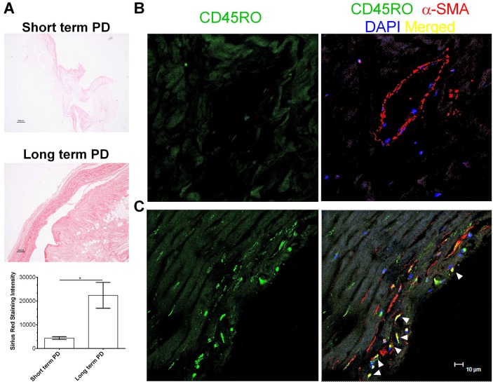
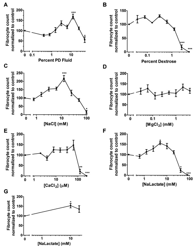
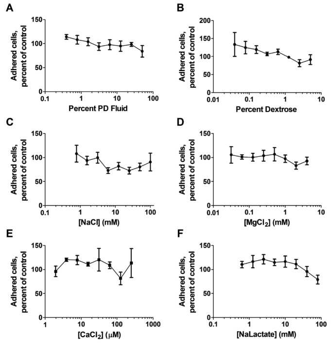
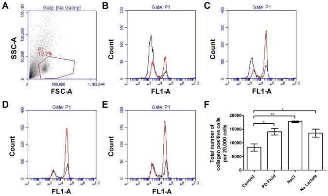
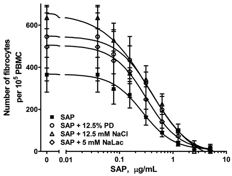
Long-term peritoneal dialysis (PD) often results in the development of peritoneal fibrosis. In many other fibrosing diseases, monocytes enter the fibrotic lesion and differentiate into fibroblast-like cells called fibrocytes. We find that peritoneal tissue from short-term PD patients contains few fibrocytes, while fibrocytes are readily observed in the peritoneal membrane of long-term PD patients. The PD fluid Dianeal (Baxter Healthcare Corporation, Deerfield, IL, USA) contains dextrose, a number of electrolytes including sodium chloride, and sodium lactate. We find that PD fluid potentiates human fibrocyte differentiation in vitro and implicates sodium lactate in this potentiation. The plasma protein serum amyloid P (SAP) inhibits fibrocyte differentiation. Peritoneal dialysis fluid and sodium chloride decrease the ability of human SAP to inhibit human fibrocyte differentiation in vitro Together, these results suggest that PD fluid contributes to the development of peritoneal fibrosis by potentiating fibrocyte differentiation.
Keywords: Peritoneal dialysis; fibrocytes; fibrosis; lactate.
Copyright © 2016 International Society for Peritoneal Dialysis.
Figures
Similar articles
-
A brief exposure to tryptase or thrombin potentiates fibrocyte differentiation in the presence of serum or serum amyloid p.J Immunol. 2015 Jan 1;194(1):142-50. doi: 10.4049/jimmunol.1401777. Epub 2014 Nov 26. J Immunol. 2015. PMID: 25429068 Free PMC article.
-
High and low molecular weight hyaluronic acid differentially regulate human fibrocyte differentiation.PLoS One. 2011;6(10):e26078. doi: 10.1371/journal.pone.0026078. Epub 2011 Oct 11. PLoS One. 2011. PMID: 22022512 Free PMC article.
-
NaCl potentiates human fibrocyte differentiation.PLoS One. 2012;7(9):e45674. doi: 10.1371/journal.pone.0045674. Epub 2012 Sep 18. PLoS One. 2012. PMID: 23029177 Free PMC article.
-
Biocompatibility of peritoneal dialysis fluid and influence of compositions on peritoneal fibrosis.Ther Apher Dial. 2006 Aug;10(4):372-9. doi: 10.1111/j.1744-9987.2006.00391.x. Ther Apher Dial. 2006. PMID: 16911191 Review.
-
New developments in peritoneal fibroblast biology: implications for inflammation and fibrosis in peritoneal dialysis.Biomed Res Int. 2015;2015:134708. doi: 10.1155/2015/134708. Epub 2015 Oct 1. Biomed Res Int. 2015. PMID: 26495280 Free PMC article. Review.
Cited by
-
Oxidative Stress in Peritoneal Dialysis Patients: Association with the Dialysis Adequacy and Technique Survival.Indian J Nephrol. 2019 Sep-Oct;29(5):309-316. doi: 10.4103/ijn.IJN_242_18. Indian J Nephrol. 2019. PMID: 31571736 Free PMC article.
-
The Development of Serum Amyloid P as a Possible Therapeutic.Front Immunol. 2018 Oct 16;9:2328. doi: 10.3389/fimmu.2018.02328. eCollection 2018. Front Immunol. 2018. PMID: 30459752 Free PMC article. Review.
References
-
- Heptinstall RH. Pathology of end-stage kidney disease. Am J Med 1968; 44:656–63. - PubMed
-
- Krediet RT, Struijk DG. Peritoneal changes in patients on long-term peritoneal dialysis. Nat Rev Nephrol 2013; 9:419–29. - PubMed
-
- Bargman JM. Advances in peritoneal dialysis: A review. Semin Dial 2012; 25:545–9. - PubMed
-
- Chan TM, Yung S. Studying the effects of new peritoneal dialysis solutions on the peritoneum. Perit Dial Int 2007; 27(Suppl 2):S87–93. - PubMed
-
- de Lima SM, Otoni A, Sabino Ade P, Dusse LM, Gomes KB, Pinto SW, et al. Inflammation, neoangiogenesis and fibrosis in peritoneal dialysis. Clin Chim Acta 2013; 421:46–50. - PubMed
MeSH terms
Substances
Grants and funding
LinkOut - more resources
Full Text Sources
Other Literature Sources
Medical
Miscellaneous
