Dysembryoplastic Neuroepithelial Tumors
- PMID: 26493957
- PMCID: PMC4696533
- DOI: 10.4132/jptm.2015.10.05
Dysembryoplastic Neuroepithelial Tumors
Abstract
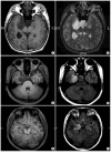
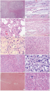
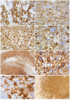
Dysembryoplastic neuroepithelial tumor (DNT) is a benign glioneuronal neoplasm that most commonly occurs in children and young adults and may present with medically intractable, chronic seizures. Radiologically, this tumor is characterized by a cortical topography and lack of mass effect or perilesional edema. Partial complex seizures are the most common presentation. Three histologic subtypes of DNTs have been described. Histologically, the recognition of a unique, specific glioneuronal element in brain tumor samples from patients with medically intractable, chronic epilepsy serves as a diagnostic feature for complex or simple DNT types. However, nonspecific DNT has diagnostic difficulty because its histology is indistinguishable from conventional gliomas and because a specific glioneuronal element and/or multinodularity are absent. This review will focus on the clinical, radiographic, histopathological, and immunohistochemical features as well as the molecular genetics of all three variants of DNTs. The histological and cytological differential diagnoses for this lesion, especially the nonspecific variant, will be discussed.
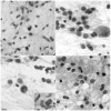
Keywords: BRAFV600E mutation; CD34; Dysembryoplastic neuroepithelial tumor; Epilepsy; Microtubule-associated protein 2.
Conflict of interest statement
No potential conflict of interest relevant to this article was reported.
Figures


Similar articles
-
Simple and complex dysembryoplastic neuroepithelial tumors (DNT) variants: clinical profile, MRI, and histopathology.Neuroradiology. 2009 Jul;51(7):433-43. doi: 10.1007/s00234-009-0511-1. Epub 2009 Feb 26. Neuroradiology. 2009. PMID: 19242688 Review.
-
A peculiar histopathological form of dysembryoplastic neuroepithelial tumor with separated pilocytic astrocytoma and rosette-forming glioneuronal tumor components.Neuropathology. 2014 Oct;34(5):491-8. doi: 10.1111/neup.12124. Epub 2014 Apr 16. Neuropathology. 2014. PMID: 24735014
-
[Dysembryoplastic neuroepithelial tumors. A benign tumor cause of partial epilepsy in young adults].Rev Neurol (Paris). 1996 Jun-Jul;152(6-7):451-7. Rev Neurol (Paris). 1996. PMID: 8944242 Review. French.
-
[Clinicopathologic features of infant dysembryoplastic neuroepithelial tumor: a case report and literature review].Beijing Da Xue Xue Bao Yi Xue Ban. 2017 Oct 18;49(5):904-909. Beijing Da Xue Xue Bao Yi Xue Ban. 2017. PMID: 29045978 Review. Chinese.
-
Dysembryoplastic neuroepithelial tumour: insight into the pathology and pathogenesis.Folia Neuropathol. 2017;55(1):1-13. doi: 10.5114/fn.2017.66708. Folia Neuropathol. 2017. PMID: 28430287 Review.
Cited by
-
Extra-temporal pediatric low-grade gliomas and epilepsy.Childs Nerv Syst. 2024 Oct;40(10):3309-3327. doi: 10.1007/s00381-024-06573-8. Epub 2024 Aug 27. Childs Nerv Syst. 2024. PMID: 39191974 Review.
-
Super T2-FLAIR mismatch sign: a prognostic imaging biomarker for non-enhancing astrocytoma, IDH-mutant.J Neurooncol. 2024 Sep;169(3):571-579. doi: 10.1007/s11060-024-04758-4. Epub 2024 Jul 12. J Neurooncol. 2024. PMID: 38995493 Free PMC article.
-
Clinicopathological features of dysembryoplastic neuroepithelial tumor: a case series.J Med Case Rep. 2023 Aug 1;17(1):327. doi: 10.1186/s13256-023-04062-1. J Med Case Rep. 2023. PMID: 37525202 Free PMC article. Review.
-
Unusual case of occipital lobe dysembryoplastic neuroepithelial tumour with GNAi1-BRAF fusion.BMJ Case Rep. 2021 Jan 27;14(1):e241440. doi: 10.1136/bcr-2020-241440. BMJ Case Rep. 2021. PMID: 33504544 Free PMC article. No abstract available.
-
Dysembryoplastic Neuroepithelial Tumor: A Case Report of A Benign Intracranial Lesion Masquerading as Seizure Disorder.Cureus. 2024 Jul 7;16(7):e64047. doi: 10.7759/cureus.64047. eCollection 2024 Jul. Cureus. 2024. PMID: 39114195 Free PMC article.
References
-
- Burneo JG, Tellez-Zenteno J, Steven DA, et al. Adult-onset epilepsy associated with dysembryoplastic neuroepithelial tumors. Seizure. 2008;17:498–504. - PubMed
-
- Daumas-Duport C, Scheithauer BW, Chodkiewicz JP, Laws ER, Jr, Vedrenne C. Dysembryoplastic neuroepithelial tumor: a surgically curable tumor of young patients with intractable partial seizures. Report of thirty-nine cases. Neurosurgery. 1988;23:545–56. - PubMed
-
- Daumas-Duport C. Dysembryoplastic neuroepithelial tumours. Brain Pathol. 1993;3:283–95. - PubMed
-
- Daumas-Duport C, Varlet P, Bacha S, Beuvon F, Cervera-Pierot P, Chodkiewicz JP. Dysembryoplastic neuroepithelial tumors: nonspecific histological forms: a study of 40 cases. J Neurooncol. 1999;41:267–80. - PubMed
Publication types
LinkOut - more resources
Full Text Sources
Other Literature Sources

