. 2015 Nov 13;350(6262):815-20.
doi: 10.1126/science.aad0217. Epub 2015 Oct 22.

MicroRNA-encoded behavior in Drosophila

Affiliations

MicroRNA-encoded behavior in Drosophila

Joao Picao-Osorio et al. Science. .

Abstract

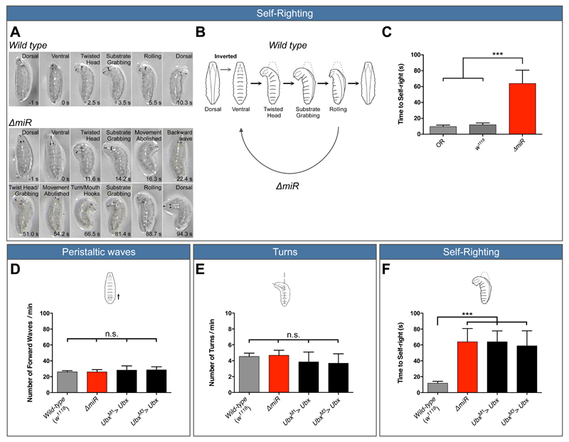
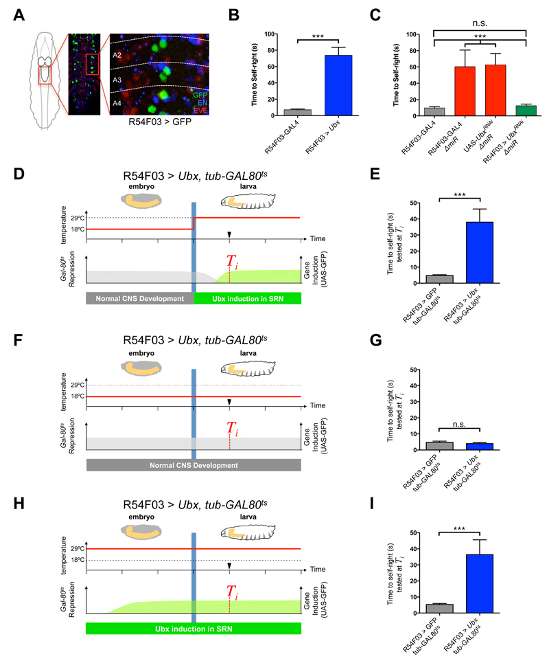
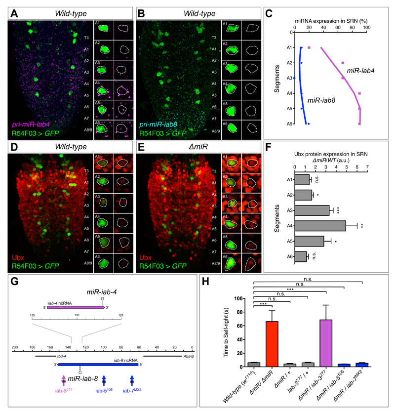
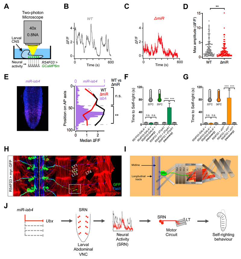
The relationship between microRNA (miRNA) regulation and the specification of behavior is only beginning to be explored. We found that mutation of a single miRNA locus (miR-iab4/iab8) in Drosophila larvae affects the animal's capacity to correct its orientation if turned upside down (self-righting). One of the miRNA targets involved in this behavior is the Hox gene Ultrabithorax, whose derepression in two metameric neurons leads to self-righting defects. In vivo neural activity analysis reveals that these neurons, the self-righting node (SRN), have different activity patterns in wild type and miRNA mutants, whereas thermogenetic manipulation of SRN activity results in changes in self-righting behavior. Our work thus reveals a miRNA-encoded behavior and suggests that other miRNAs might also be involved in behavioral control in Drosophila and other species.


Figures

Figure 1
Figure 1. Both, removal of miR-iab4/iab8 and over-expression of Ubx disrupt a specific larval locomotor behaviour: self-righting
(A, B) Description of larval self-righting behaviour. (A) Time-lapses of larval self-righting behaviour. (A top) Wild type larvae were placed in an inverted position (ventral up), twisted their heads, grabbed the substrate with the mouth hooks and rolled their bodies onto their ventral surface (dorsal up). In contrast, (A bottom) ∆miR larvae displayed problems in self-righting their bodies. (B) Diagram of the self-righting behavioural response. (C) Quantification of the time required for the successful completion of the self-righting behaviour (mean ± SEM; N =27 to 29 larvae per genotype) in the two wild type controls (OR, w1118, light and dark grey respectively) and ∆miR larvae (red). (D-F) Quantification of larval behaviour in Ubx overexpression lines (UbxM1>Ubx and UbxM3>Ubx). Quantification of (D) number of forward peristaltic waves per minute, (E) larval turning per minute and (F) time to self-right in: wild-type (w1118, grey), ∆miR (red), UbxM1> Ubx and UbxM3> Ubx (black) (mean ± SEM; N =15 to 29 larvae per genotype). A non-parametric Mann-Whitney U test was performed to compare treatments; p > 0.05 (non-significant; n.s); p < 0.001 (***).
Figure 2
Figure 2. miRNA-dependent Ubx regulation in SRN cells underlies SR behaviour
(A) R54F03-GAL4 expression (GFP, green) in the larval abdominal CNS (Even-skipped protein in red; Engrailed protein in blue; A2 refers to abdominal segment 2). (B) Artificial increase of Ubx expression in two metameric neurons driven by the R54F03-GAL4 promoter (mean ± SEM; N = 60 per genotype). (C) Artificial decrease of Ubx expression with UbxRNAi within SRN cells in ∆miR larvae (R54F03-GAL4, ∆miR/ UAS-UbxRNAi, ∆miR, green) (mean ± SEM; N = 20 to 23 per genotype). (D-I) Conditional increase of Ubx expression during embryonic and early larval development with tub-Gal80ts (Gal80ts represses GAL4 activity at 18°C) within SRN cells: R54F03 > Ubx, tub-GAL80ts (UAS-Ubx /+ ; R54F03-GAL4/ tub-Gal80ts). Controlled increase of Ubx expression in SRN cells in early larvae (D and E; mean ± SEM; N = 20 per genotype) and from mid-embryogenesis to early larvae (H and I; mean ± SEM; N = 15 per genotype). (F and G) Repressed increase of Ubx expression in SRN cells throughout embryogenesis and early larvae (mean ± SEM; N = 15 per genotype). A non-parametric Mann-Whitney U test was performed to compare treatments; p>0.05 (non-significant; n.s.); p<0.001 (***).
Figure 3
Figure 3. Regulation of Ubx protein expression in SRN cells by miR-iab4/iab8
(A) Wild type expression of precursor miR-iab4 transcripts (RNA-FISH, magenta) in SRN cells (R54F03>GFP, green) of the ventral nerve cord (VNC) of first-instar Drosophila larvae. (B) Wild type expression of precursor miR-iab8 transcripts (RNA-FISH, cyan) in SRN cells (R54F03>GFP, green) of the VNC of first-instar Drosophila larvae. (C) Percentage of SRN cells expressing miR-iab4 (purple, square) and miR-iab8 (blue, triangle) precursors across A1 to A6 (N = 10). (D and E) Ubx protein expression (red) in SRN cells of wild type (D) and ∆miR (E) first-instar larvae VNCs. (F) Quantification of Ubx protein expression ratio of ∆miR over wild type within the SRN cells (red) by fluorescent intensity (N = 8 per genotype; arbitrary units, a.u.). (G) Diagram of a sub-region of the bithorax complex based on (13) showing iab-4 (purple) and iab-8 (blue) non-coding RNAs (ncRNA), and rearrangement breakpoints affecting miR-iab-4 (iab-3277, purple) and miR-iab-8 (iab-5105 and iab-7MX2, blue). (H) Genetic complementation tests to determine the involvement of miR-iab4 or miR-iab8 in SR behaviour using trans-heterozygote larvae for ∆miR and different chromosomal rearrangement breakpoints that disrupt the bithorax complex (mean ± SEM; N = 17 to 20 per genotype). A non-parametric Mann-Whitney U test was performed to compare treatments; p>0.05 (non-significant; n.s.); p < 0.05 (*); p < 0.01 (**); p<0.001 (***).
Figure 4
Figure 4. ∆miR mutants have abnormal patterns of neural activity in SRN cells
(A) Schematic of the lateral larval CNS expressing GCaMP6m in SRN cells (R54F03> GCaMP6m, green) imaged in a two-photon microscope. (B) Examples of spontaneous activity recorded over 10 mins from wild type (WT: UAS-GCaMP6m/+; R54F03-GAL4/+) and (C) ∆miR mutants (UAS-GCaMP6m/+; R54F03-GAL4, ∆miR/∆miR) in SRN cells. (D) Maximum amplitude of spontaneous activity in SRN cells: WT (median ∆F/F=1.91; N = 120) and in ∆miR mutants (median ∆F/F=1.27; N = 115) (** p<0.01, Mann-Whitney U test). (E) Expression pattern of miR-iab4 (purple) and DAPI (blue) in the VNC of a freshly hatched larva (left panel). Median ∆F/F in SRN cells of WT (black line) and ∆miR (red line) larval VNCs, and relative expression of miR-iab4 (purple) along the anterior-posterior (A-P) axis. Median ∆F/F of WT (median of 2.132, n=73) and ∆miR (median of 1.122, n=68) in regions of high miR-iab4 expression (** p<0.01, Mann-Whitney U test). Regions of low miR-iab4 expression have a median ∆F/F of 1.763 in WT (N = 47) and 1.749 (N = 47) in ∆miR specimens (n.s., p>0.05; Mann-Whitney U test). (F and G) Thermogenetic manipulation of neural activity in SRN cells. Activation (F, R54F03>dTrpA1) and inhibition (G, R54F03>shits) of SRN neural activity (*** p<0.0001) [29°C (green) for activation (H) and 36°C (orange) for inhibition (I)]. (H) Wild-type motor axonal projections of SRN cells (UAS-myr::GFP/ UAS-myr::GFP; R54F03-GAL4/ R54F03-GAL4, green) into muscles (phalloidin, red) lateral transverse 1 and 2 (LT1 and LT2) in late embryos (stage 17)(Fasciclin II, FASII, blue).). Scale bars (white bars) represent 10μm (I) Diagram of SRN cells projecting to the LT1 and LT2 muscles. (J) A model that summarises the data reported in this study. Mutation of miR-iab4 (left) leads to Ubx de-repression in the SRN node affecting SRN neural activity patterns and triggering an anomalous self-righting behaviour (right).

References

    1. Li X, Jin P. Nat Rev Neurosci. 2010;11:329–338. - PubMed
    1. Sun AX, Crabtree GR, Yoo AS. Curr Opin Cell Biol. 2013;25:215–221. - PMC - PubMed
    1. Thomsen S, Azzam G, Kaschula R, Williams LS, Alonso CR. Development. 2010;137:2951–2960. - PubMed
    1. Reed HC, et al. Genetics, genetics. 2010;109:112086.
    1. de Navas LF, et al. Development. 2011;138:107–116. - PubMed

Publication types