. 2015 Nov 10;112(45):E6185-94.
doi: 10.1073/pnas.1519040112. Epub 2015 Oct 22.

Fetal endocannabinoids orchestrate the organization of pancreatic islet microarchitecture

Affiliations

Fetal endocannabinoids orchestrate the organization of pancreatic islet microarchitecture

Katarzyna Malenczyk et al. Proc Natl Acad Sci U S A. .

Abstract

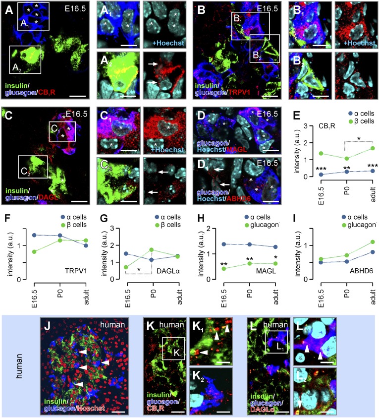
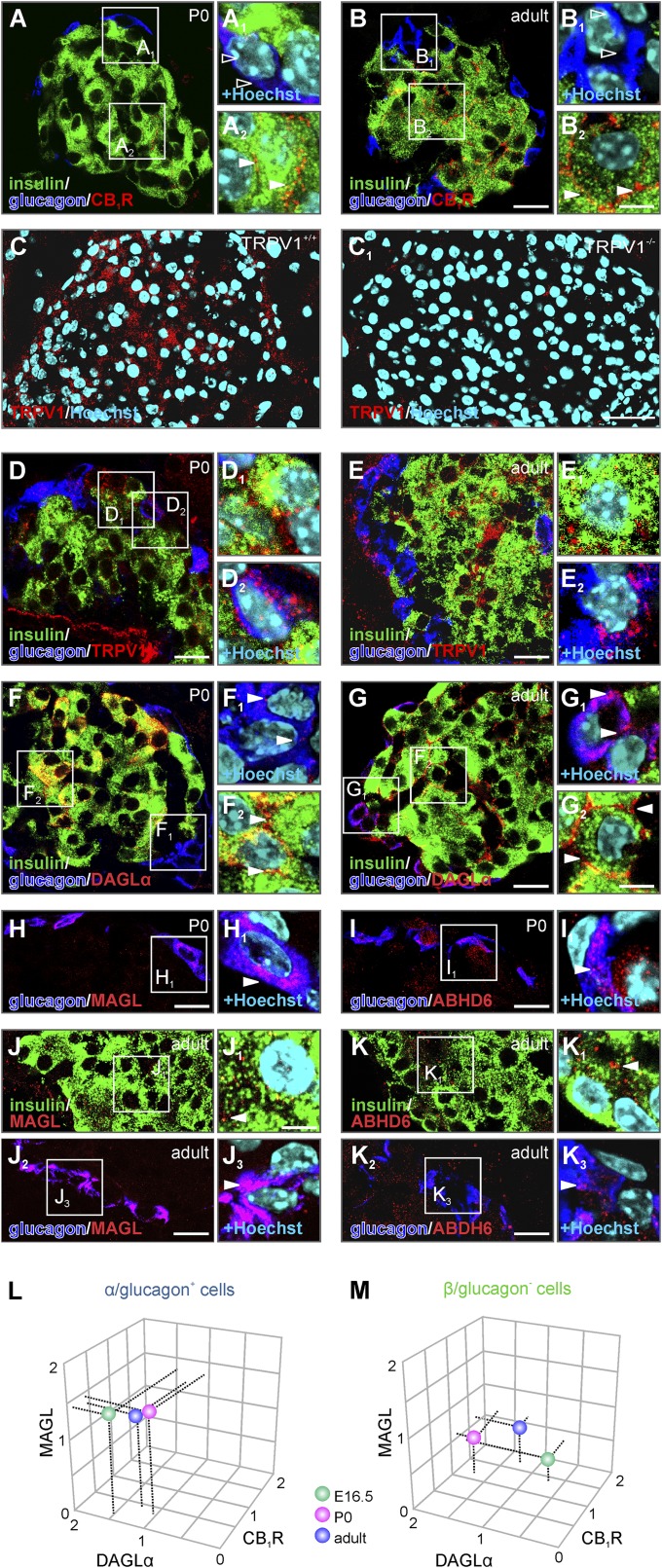
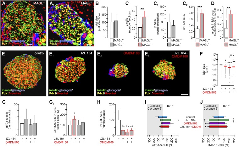
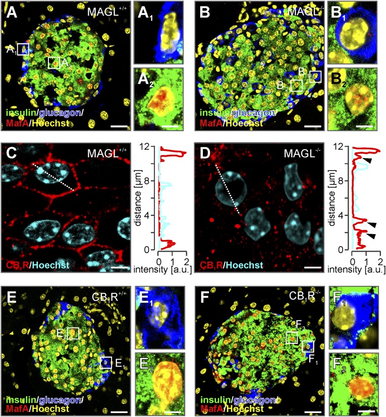
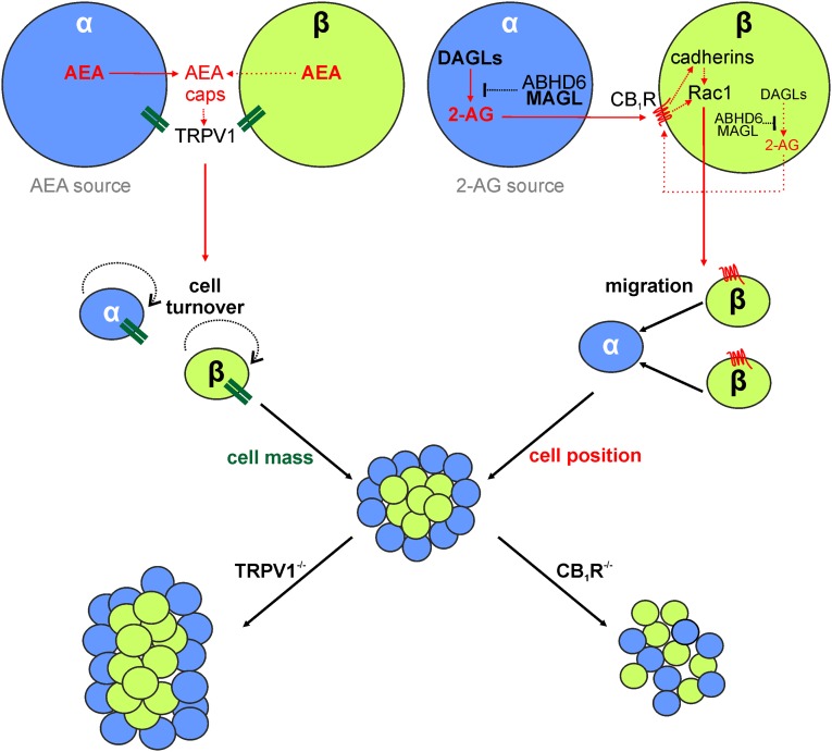
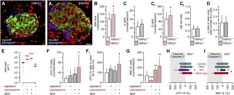
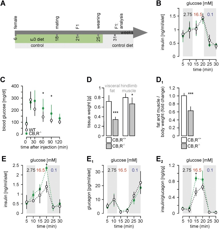
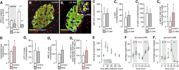
Endocannabinoids are implicated in the control of glucose utilization and energy homeostasis by orchestrating pancreatic hormone release. Moreover, in some cell niches, endocannabinoids regulate cell proliferation, fate determination, and migration. Nevertheless, endocannabinoid contributions to the development of the endocrine pancreas remain unknown. Here, we show that α cells produce the endocannabinoid 2-arachidonoylglycerol (2-AG) in mouse fetuses and human pancreatic islets, which primes the recruitment of β cells by CB1 cannabinoid receptor (CB1R) engagement. Using subtractive pharmacology, we extend these findings to anandamide, a promiscuous endocannabinoid/endovanilloid ligand, which impacts both the determination of islet size by cell proliferation and α/β cell sorting by differential activation of transient receptor potential cation channel subfamily V member 1 (TRPV1) and CB1Rs. Accordingly, genetic disruption of TRPV1 channels increases islet size whereas CB1R knockout augments cellular heterogeneity and favors insulin over glucagon release. Dietary enrichment in ω-3 fatty acids during pregnancy and lactation in mice, which permanently reduces endocannabinoid levels in the offspring, phenocopies CB1R(-/-) islet microstructure and improves coordinated hormone secretion. Overall, our data mechanistically link endocannabinoids to cell proliferation and sorting during pancreatic islet formation, as well as to life-long programming of hormonal determinants of glucose homeostasis.

Keywords: G protein-coupled receptor; cell adhesion; diabetes; migration; proliferation.


Conflict of interest statement

The authors declare no conflict of interest.

Figures

Fig. 1.
Fig. 1.
Spatial segregation of CB1R and DAGLα in the fetal endocrine pancreas. (A–A2) CB1R immunoreactivity was seen in insulin+/β cells (arrows) but not in glucagon+/α cells (asterisks) at E16.5. (B–B2) TRPV1 immunosignals were found in both glucagon+/α cells and insulin+/β cells. (C–C2) In contrast, glucagon+/α cells (asterisks) were the primary sites of DAGLα expression at this developmental stage. (D) Likewise, MAGL was predominantly localized to glucagon+/α cells (asterisks). (D1) ABHD6 immunoreactivity localized to both glucagon+/α (asterisks) and glucagon cells (arrows). Rectangles indicate the position of Insets. Histochemical data from newborn and adult mice are shown in Fig. S2. (E and F–I) Semiquantitative analysis of CB1R, TRPV1, DAGLα, MAGL, and ABHD6 immunosignals at successive developmental stages. Statistical comparisons were performed by nonparametric Wilcoxon’s rank-sum test, n = 10 clusters or islets per group. Data are expressed as medians; ***P < 0.001, **P < 0.01, *P < 0.05. (J–L2) Glucagon+/α and insulin+/β cell distribution in human pancreatic islets. (J) Note that glucagon+/α cells are positioned in pancreatic islet core (arrowheads). CB1R (K–K2) and DAGLα immunosignals (L–L2) predominated in insulin+/β cells (arrowheads) and glucagon+/α cells, respectively. Hoechst 33342 was used as nuclear counterstain. (Scale bars: J, 25 μm; A, B, C, K, and L, 10 μm; A1, A2, B1, B2, C1, C2, D, D1, K1, K2, L1, and L2, 5 μm.)
Fig. S1.
Fig. S1.
CB1R, TRPV1, DAGLα, MAGL, and ABHD6 distribution in the endocrine pancreas of neonatal (P0) and adult mice. (A–B2) CB1R immunoreactivity decorated the majority of insulin+ β cells at both ages. In contrast, CB1R expression in glucagon+ α cells remained below detection threshold in the endocrine pancreas at both P0 and adulthood. (C and C1) Histochemical control demonstrating the specificity of rabbit anti-TRPV1 in the pancreas of adult WT and TRPV1−/− mice. The lack of residual immunolabeling in TRPV1−/− tissue supports the specificity of the immunoreagent used. (D–E2) TRPV1s were detected in insulin+ β cells and glucagon+ α cells at both ages. (F–G2) DAGLα was found in both insulin+ and glucagon+ cells by birth and postnatally. (H, H1, and J–J3) MAGL primarily localized to glucagon+ cells. However, some residual immunosignal was observed in glucagon (likely β) cells. (I, I1, and K–K3) ABHD6 was uniformly distributed in neonatal and adult pancreatic islets. Hoechst 33342 was used as nuclear counterstain. (Scale bars: C and C1, 15 μm, 40 µm; Insets, 5 μm.) (L and M) The 3D rendering of a minimal 2-AG signaling cassette showed developmental rearrangements in glucagon (likely β) cells, with limited changes in glucagon+/α cells, suggesting that β cells are an eCB-responsive cell contingent.
Fig. 2.
Fig. 2.
The cellular organization of pancreatic islets is controlled by 2-AG signaling. (A and A1) Glucagon+/α and insulin+/Pdx1+/β cell distribution in pancreatic islets from adult MAGL−/− mice relative to WT littermates. Quantitative analysis was performed in n ≥ 3 animals per genotype. (B) Genetic disruption of MAGL did not affect pancreatic islet size. (C and C1) However, pancreatic islets from MAGL−/− mice contained significantly more α cells whereas the number of β cells remained unchanged. (C2) Thus, the ratio of α/β cells significantly increased in pancreatic islets from MAGL−/− mice. (D) Genetic manipulation induced incomplete α/β cell segregation with α cells seen in pancreatic islet cores. (E–E3) JZL184, an MAGL inhibitor that significantly increases extracellular 2-AG availability (2), as well as OMDM188, a DAGL inhibitor (39), reduced pseudoislet assembly. Hoechst 33342 was used as nuclear counterstain (pseudocolored in red). (Scale bars: 25 µm; Insets, 5 µm.) (F) Both JZL184 and OMDM188 alone or in combination significantly reduced the size of pseudoislets. (G) Neither treatment affected the number of CB1R αTC1-6 cells. Nevertheless, JZL184 induced significant αTC1-6 spread into the pseudoislets’ core (G1). (H) Both JZL184 and OMDM188 significantly reduced the number of INS-1E/β cells in pseudoislets. (I and J) The rate of apoptosis and cell proliferation, measured by cleaved caspase-3 and Ki67 immunoreactivity, respectively, in αTC1-6 and INS-1E cells cocultured in vitro. Representative images are shown in Fig. S7. (I) JZL184 but not OMDM188 increased αTC1-6 cell proliferation and decreased the rate of cell death. (J) Quantitative analysis of the rate of apoptosis and proliferation in INS-1E cells revealed increased cell turnover by JZL184. OMDM188 alone or in combination with JZL184 significantly augmented the number of apoptotic INS-1E cells. Data are expressed as means ± SD; n = 30 islets per genotype (B–D), n ≥ 100 islets per group (F), n = 10 islets per group (G–H), n ≥ 300 cells per group (I and J) were analyzed in triplicate experiments, ***P < 0.001, **P < 0.01, *P < 0.05 [Student’s t test (B–D) or post hoc pairwise comparisons, one-way ANOVA].
Fig. S2.
Fig. S2.
β and α cells retain lineage characteristics in MAGL−/− and CB1R−/− mice. (A–B2) MafA immunosignal was detected in β but not α cells in both adult MAGL−/− mice and their WT littermates. (C and D) Intracellular CB1Rs, suggesting receptor desensitization, were observed in β cells of MAGL−/− but not MAGL+/+ mice. (E–F2) MafA immunosignals were present in β but not α cells in pancreata of adult CB1R−/− mice and WT littermates. Hoechst 33342 was used as nuclear counterstain (pseudocolored in yellow). (Scale bars: 25 µm; Insets, 2.5 µm.)
Fig. S3.
Fig. S3.
Schema of the proposed role of cell type-specific eCB signaling at CB1Rs and/or TRPV1s in the regulation of pancreatic islet size and cell sorting. The α cells are the main source of locally acting AEA and 2-AG. Activation of TRPV1 signaling in pancreatic α and/or β cells [AEA, capsaicin (caps)] increases α and β cell turnover, thus limiting islet size. Accordingly, TRPV1 inhibition and genetic deletion enlarges pancreatic islets. In turn, DAGLs in α cells produce 2-AG, which uses a paracrine signaling mechanism to activate CB1Rs in β cells. CB1R might also be activated cell-autonomously by 2-AG through DAGLs residing in β cells. CB1R signaling affects adhesion signaling (cadherins), cell motility (Rac1), promoting α- and β-cell segregation into a core-mantle structure. Inhibition of CB1R signaling (e.g., CB1R−/−) impairs cells segregation, resulting in the formation of mixed islets.
Fig. S4.
Fig. S4.
Molecular determinants of cell motility in α and β cells. (A–C) Rac1 was found in both α and β cells at E16.5 (A); but only in β cells at P0 (B) and by adulthood (C). (D–F1). Doublecortin (DCX), an intermediate filament expressed by migrating neurons in the nervous system (34) and also detected in the pancreas (33), was predominantly visualized in glucagon+ (α) cells in both pre- and postnatal pancreatic islets. (G–I1) In turn, insulin+ (β) cells expressed DCX at markedly lower levels. Hoechst 33342 was used as nuclear counterstain. (Scale bars: Insets, 5 µm; C, D, and G, 10 µm; E and H, 20 µm; F and I, 40 µm.)
Fig. S5.
Fig. S5.
Molecular determinants of endocannabinoid signaling in αTC1-6 cells. (A) Reverse-transcription PCR products (arrowheads) of select receptor and enzyme components of 2-AG and AEA signaling. Tissue samples from the cerebral cortex, cerebellum, and spleen served as positive controls. Samples without reverse transcriptase (RT) were used as negative controls. αTC1-6 cells did not express CB1R receptor mRNA at detectable levels (open arrowhead). (B) Western analysis of a select battery of molecular markers in αTC1-6 cells (86). Arrows point to immunoreactive protein bands at the calculated molecular mass of each target. Two MAGL isoforms are shown. (C and D) MAGL and DAGLα localization in αTC1-6 cells by fluorescence microscopy. Hoechst 33342 was used as nuclear counterstain. (Scale bar: 5 µm.) (E and E1) αTC1-6 contain and release significantly more 2-AG than INS-1E cells whereas the level of AEA in cell pellets is less drastically different from that in INS1-E cells. Data are expressed as means ± SD from triplicate experiments. *P < 0.05 (pairwise comparisons).
Fig. S6.
Fig. S6.
Pseudoislets, α/β cell aggregates, as a model of pancreatic islets in vitro. (A) Suspension coculture of INS-1E and αTC1-6 cells (2:1) for 48 h results in the formation of cell aggregates (pseudoislets) as revealed by phase-contrast microscopy. (B) Confocal microscopic orthogonal image stacks of glucagon and insulin-labeled pseudoislets demonstrate that cell aggregates retain the 3D core-mantle morphology of native pancreatic islets. (C) Vinculin was used to detect sites of cell adhesion. (D–F) Simultaneous measurement of insulin and glucagon secretion from pseudoislets pretreated with JZL184 (200 nM) alone or in combination with OMDM188 (100 nM) for 24 h. JZL184 pretreatment increased both basal insulin and glucagon release. OMDM188 pretreatment reduced hormone release and occluded the effect of JZL184. (F) Insulin/glucagon ratio revealed a significant shift toward insulin release when analyzing coordinated hormone release from JZL184-exposed pseudoislets. Hoechst 33342 was used as nuclear counterstain. (Scale bars: A, 75 μm; B, 40 μm; C, 25 μm.) Data are expressed as means ± SD; n ≥ 10 from triplicate experiments. *P < 0.05 (pairwise comparisons, one-way ANOVA).
Fig. S7.
Fig. S7.
Effect of CB1R and TRPV1 signaling on α- and β-like cell proliferation and survival in vitro. Representative images of αTC1-6 cells (glucagon+) mixed with INS-1E cells (glucagon) in vitro. Mixed cultures were exposed to the drugs indicated. (A–A3) JZL184 increased the proliferation of both αTC1-6 and INS-1E cells, as revealed by quantitative analysis of Ki67 immunoreactivity (40). (B–B3) In contrast, OMDM188 alone or in combination with JZL184 augmented INS-1E cell death (as shown by cleaved caspase-3 immunoreactivity) without affecting αTC1-6 survival. Quantitative data are shown in Fig. 2 H and I. (C–C3) CB1R manipulation selectively affected INS-1E cells proliferation and (D–D3) survival. Data from quantitative analysis are shown in Fig. 4 H and I. (E–E3) Capsazepine significantly slowed the proliferation of both αTC1-6 and INS-1E cells. (F–F3) Moreover, capsazepine decreased INS-1E cell death, as indicated by the significantly reduced number of cleaved caspase-3+ cells, without affecting αTC1-6 cell survival. Quantitative data are shown in Fig. 5 H and I. Hoechst 33342 was used as nuclear counterstain (pseudocolored in red). (Scale bars: 15 µm.)
Fig. 3.
Fig. 3.
The 2-AG signals modulate adhesion signaling in pancreatic islets. (A–C1) MAGL knockout alters cell adhesion in the adult endocrine pancreas as revealed by E-cadherin immunostaining (A and A1). E-cadherin immunoreactivity decreased, particularly in the cytoplasm of β cells (B and B1). In contrast, we found increased membrane-localized E-cadherin in α cells of MAGL−/− mice (C and C1). Hoechst 33342 was used as nuclear counterstain. (Scale bars: A and A1, 5 μm; B–C1, 2.5 μm.) (D–F) Quantitative analysis of the intensity and distribution of E-cadherin immunosignals within pancreatic islets (indiscriminate; D), α (E), and β cells (F) of WT and MAGL−/− mice. Data are expressed as means ± SD; n = 10 islets per genotype (D), n = 30 cells per genotype (E and F), n = 10 islets per group were analyzed in triplicate. ***P < 0.001, **P < 0.01, *P < 0.05 (Student’s t test). cyto, cytoplasmic; membr, membraneous; perinuc, perinuclear.
Fig. 4.
Fig. 4.
CB1R activity orchestrates α and β cell sorting in fetal endocrine pancreas. (A and A1) Glucagon+/α and insulin+/Pdx1+/β cell distribution in pancreatic islets from adult CB1R−/− mice relative to WT littermates. Quantitative analysis was performed in n ≥ 3 animals per genotype. (B) Genetic disruption of CB1R activity did not affect pancreatic islet size. (C and C1) However, pancreatic islets from CB1R−/− mice contained significantly more α cells while leaving the β cell pool unchanged. (C2) Thus, the ratio of α/β cells significantly increased in pancreatic islets from CB1R−/− mice. (D) Moreover, CB1R knockout induced incomplete α/β cell segregation with α cells residing in pancreatic islet core. (E) AEA increased the size of pseudoislets formed. O-2050 antagonized this effect by sorting αTC1-6 and INS-1E cells to separate clusters. (F) AEA and O-2050 increased the number of αTC1-6 cells in individual pseudoislets, including their localization within the pseudoislets’ core (F1). (G) INS-1E cell numbers were not altered by either AEA or O-2050 alone. However, combined treatment led to the formation of small pseudoislets primarily consisting of only one cell type. (H and I) Quantitative analysis of the rates of apoptosis and proliferation for αTC1-6 and INS-1E cells cultured in the presence of the ligands indicated. Representative images are shown in Fig. S7. Note that CB1R modulation primarily affected INS-1E cell turnover. Data are expressed as means ± SD; n = 30 islets per genotype (B–D), n ≥ 100 islets per group (E), n = 10 islets per group (F–G), n ≥ 300 cells per group (H and I) were analyzed in triplicate experiments, ***P < 0.001, **P < 0.01, *P < 0.05 [Student’s t test (B–D) or pairwise comparisons/one-way ANOVA].
Fig. S8.
Fig. S8.
CB1R activity orchestrates α and β cell sorting in fetal endocrine pancreas whereas TPRV1 signaling controls the size of pancreatic islets. (A–A3) Anandamide (AEA)-induced CB1R activation resulted in ectopic αTC1-6 cell localization. In contrast, O-2050, a CB1R antagonist, alone increased the number of αTC1-6 recruited during the process of “mantle formation” (arrowheads) in vitro. Moreover, O-2050, when combined with AEA, led to the segregation of pseudoislets predominantly containing either INS-1E (open arrowheads) or αTC1-6 cells (arrowheads). Quantitative data are shown in Fig. 4 EG. (B–B3) URB597 (100 nM, FAAH inhibitor) neither increased pseudoislet size significantly nor affected αTC1-6 and INS-1E cells numbers. Instead, it increased the number of αTC1-6 cells within the pseudoislets’ core (B3). (C–C3) Representative images of pseudoislets formed in the presence of capsaicin (300 nM, TRPV1 agonist), and capsazepine (cpz; 10 μM, TRPV1 antagonist) alone or in combination with AEA (10 μM, endogenous TRPV1 agonist). Quantitative data are shown in Fig. 5 EG. (D–D3) AMG 9810 (1 μM, TRPV1 antagonist) (82) increased pseudoislet size through elevating INS-1E cell numbers. In contrast, AEA signaling through cannabinoid receptors in the presence of AMG 9810 induced αTC1-6 cell recruitment and translocation toward pseudoislet cores. (Scale bars: A–A3, 25 µm; C–C3, 40 µm.) Data are expressed as means ± SD; n = 10 pseudoislets per group were analyzed in triplicate experiments, ***P < 0.001, **P < 0.01, *P < 0.05 [Student’s t test (B–B3) or pairwise comparisons/one-way ANOVA].
Fig. 5.
Fig. 5.
TPRV1 signaling controls the size of pancreatic islets. (A and A1) Glucagon+/α and insulin+/β cell distribution in pancreatic islets from adult TRPV1−/− mice relative to WT littermates. Quantitative analysis was performed in n ≥ 3 animals per genotype. (B) Genetic disruption of TRPV1 increased pancreatic islet size. (C and C1) Although the number of α cells in pancreatic islets from TRPV1−/− mice remained unaffected, they contained significantly more β cells. (C2 and D) Neither the ratio of α/β cells nor their relative positions changed. (E) Capsaicin (300 nM, TRPV1 agonist) reduced, whereas capsazepine (cpz) (10 μM, TRPV1 antagonist) increased pseudoislet size. (F and F1) TRPV1 modulation alone did not affect the number or positioning of αTC1-6 cells. In contrast, AEA (10 μM, endogenous TRPV1 and CB1R agonist) signaling through cannabinoid receptors in the presence of capsazepine induced αTC1-6 cell recruitment. Thus, AEA distinguished CB1R and TRPV1-selective mechanisms. (G) Capsaicin significantly abrogated whereas capsazepine alone or in combination with AEA increased the number of INS-1E cells. (H and I) Quantitative assessment of proliferation and apoptosis for αTC1-6 and INS-1E cells. Representative images are shown in Fig. S7. Capsazepine reduced the rate of cell proliferation in both cell lines, as well as reduced INS-1E cell death in vitro. AEA reversed the antiproliferative effect of capsazepine. Hoechst 33342 was used as nuclear counterstain (pseudocolored in red). (Scale bar: 25 µm.) Data are expressed as means ± SD; n = 20 islets per group (B–D). n ≥ 100 islets per group (E), n = 10 pseudoislets per group (F–G), n ≥ 300 cells per group (H and I) from triplicate experiments, ***P < 0.001, **P < 0.01, *P < 0.05 [pairwise comparisons after one-way ANOVA or Student’s t test (B–D)].
Fig. S9.
Fig. S9.
Dietary enrichment in ω3-PUFA during development, as well as genetic ablation of CB1R expression improves secretory responses in pancreatic islets. (A) Schema of experimental design. (B) In primary mouse islets isolated from adult mice (10 wk) exposed to ω3-PUFA–enriched diet during in utero development and until weaning, basal and glucose-stimulated release of insulin was unaffected. (C) Glucose intolerance (slowed glucose clearance) in CB1R−/− mice (n > 6 per group). (D and D1) Mass of visceral fat and hind limb muscle dissected from CB1R−/− mice is significantly lower than in WTs. These changes remain when calculated as the ratio of fat and muscle/total body weight. (E and E1) In primary mouse islets isolated from control and CB1R−/− mice, 16.5 mM glucose-stimulated release of insulin was increased whereas glucagon secretion was unaffected. (E2) Therefore, the correlated insulin/glucagon ratio was significantly increased in the presence of high glucose. Data are expressed as means ± SD; n = 30 islets per group from triplicate experiments, ***P < 0.001, *P < 0.05 [Student’s t test (D and D1) or pair-wise comparisons after one-way ANOVA].
Fig. 6.
Fig. 6.
Incomplete α/β cell sorting leads to improved secretory responses in pancreatic islets. (A) ω3 polyunsaturated fatty acid (PUFA)-enriched diet resulted in decreased AEA levels in the blood of offspring as determined by mass spectrometry. (B and B1) Pancreatic islet morphology in animals fed on normal (B) vs. ω3-PUFA–enriched diets until weaning (B1). Localization of α cells in ω3-PUFA–fed mice (Inset, arrows). Hoechst 33342 was used as nuclear counterstain (pseudocolored in red). (Scale bars: B1, 40 μm; Inset, 20 μm.) (C–C3) Neither the size of individual islets (C) nor their α/β cell composition (C1 and C2) was quantitatively altered. Instead, α cells resided in the islet core (>10 μm from the surface; C3). (D–D3) Quantitative assessment of pseudoislets formed in the presence of docosahexaenoic acid (DHA, 10 µM). DHA increased pseudoislet size (D) but not the numbers of αTC1-6 (D1) and INS-1E (D2) cells forming these clusters. (D3) Instead, the number of αTC1-6 cells residing in the psudoislet core was significantly increased. (E) Mice prenatally exposed to ω3-PUFAs showed improved glucose tolerance (n > 6 per group). (F) In primary mouse islets isolated from ω3-PUFA–fed mice, basal release of glucagon was decreased. (F1) Moreover, the correlated insulin/glucagon ratio was significantly increased upon high glucose. n ≥ 3 animals per group were quantitatively assessed. Data are expressed as means ± SD; n = 30 islets per group or n = 10 pseudoislets per group from triplicate experiments, **P < 0.01, *P < 0.05 [Student’s t test (A–D3) or pairwise comparisons after one-way ANOVA].

Similar articles

Cited by

References

    1. Matias I, Di Marzo V. Endocannabinoids and the control of energy balance. Trends Endocrinol Metab. 2007;18(1):27–37. - PubMed
    1. Malenczyk K, et al. CB1 cannabinoid receptors couple to focal adhesion kinase to control insulin release. J Biol Chem. 2013;288(45):32685–32699. - PMC - PubMed
    1. Li C, et al. Cannabinoid receptor agonists and antagonists stimulate insulin secretion from isolated human islets of Langerhans. Diabetes Obes Metab. 2011;13(10):903–910. - PubMed
    1. Vilches-Flores A, Hauge-Evans AC, Jones PM, Persaud SJ. Chronic activation of cannabinoid receptors in vitro does not compromise mouse islet function. Clin Sci (Lond) 2013;124(7):467–478. - PubMed
    1. Li C, et al. Expression and function of monoacylglycerol lipase in mouse β-cells and human islets of Langerhans. Cell Physiol Biochem. 2012;30(2):347–358. - PubMed

Publication types

LinkOut - more resources