Review
. 2015 Sep;30(3):97-106.
doi: 10.1053/j.tcam.2015.07.008. Epub 2015 Jul 9.

Hyphema: Considerations in the Small Animal Patient

Affiliations
Review

Hyphema: Considerations in the Small Animal Patient

Mary Rebecca Telle et al. Top Companion Anim Med. 2015 Sep.

Abstract

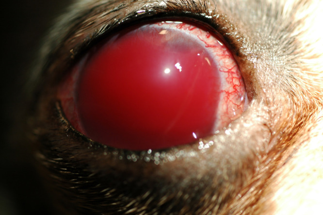
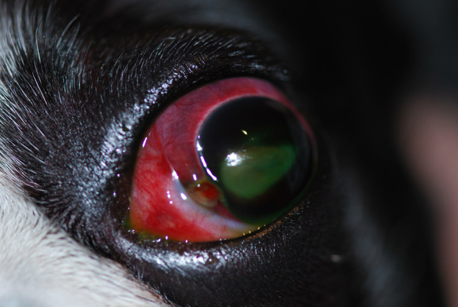
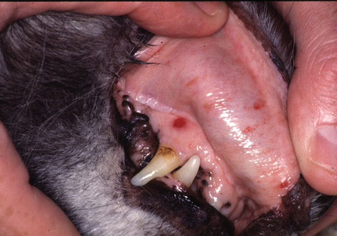
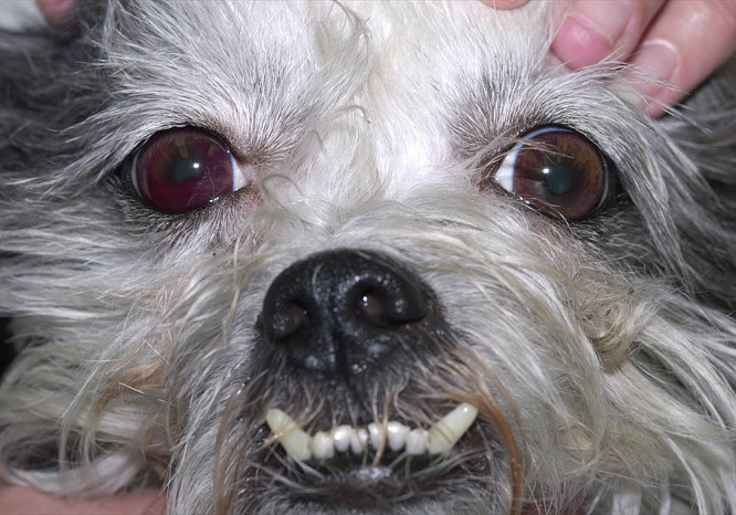
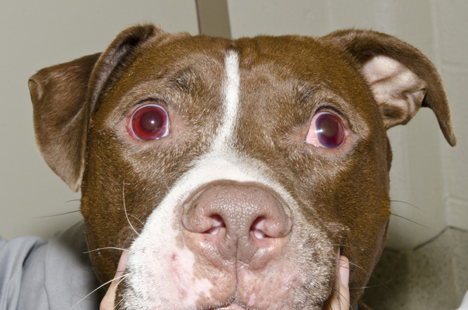
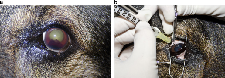
Classification, diagnosis, and treatment of hemorrhage into the anterior chamber of the eye, or hyphema, can be a challenging and frustrating process for many practitioners, especially in emergency situations. This review outlines an inclusive list of causes, diagnostics, and treatments for traumatic and nontraumatic hyphema in both canine and feline patients. The review is tailored to small animal practitioners, especially in emergency practice, and is designed to provide concise but thorough descriptions on investigating underlying causes of hyphema and treating accordingly.

Keywords: bleeding; canine; emergency; feline; hemorrhage; hyphema.


Figures

Fig. 1
Fig. 1
Anterior chamber containing unclotted blood. Unclotted hyphema often indicates continued bleeding or lack of the ability to form a clot. (Image courtesy of Ellen Belknap.)
Fig. 2
Fig. 2
An “8-ball” appearance of hyphema following vehicular trauma. The hyphema has a blue or black appearance, which is typical of clot formation and may lead to secondary glaucoma. The cornea is perforated at the medial limbus and there is severe exophthalmos due to retrobulbar and subconjunctival hemorrhage. A corneal ulcer has developed owing to globe exposure.
Fig. 3
Fig. 3
Petechiae present in a patient with immune-mediated thrombocytopenia. It is important to perform a full physical examination on any patient presenting for hyphema because subtle findings of petechiae or ecchymoses may prompt further investigation of underlying systemic coagulation disorders. (Image courtesy of Andrew Mackin.)
Fig. 4
Fig. 4
Shih Tzu with immune-mediated thrombocytopenia. The right eye has hyphema and aqueous flare. Platelet disorders are a common cause of hyphema especially if vasculitis is present or the platelets are extremely low. (Image courtesy of Andrew Mackin.)
Fig. 5
Fig. 5
Hyphema present in association with multicentric lymphoma and anterior uveitis in a 6-year-old pit bull terrier. Aqueous flare with a small amount of hyphema is present in both eyes.
Fig. 6
Fig. 6
This is the same patient as seen in Fig 5. All palpable lymph nodes were enlarged and firm.
Fig. 7
Fig. 7
Doppler blood pressure measurement in a 13-year-old domestic shorthaired cat. If hyphema is seen, blood pressure measurement is indicated as part of a thorough examination.
Fig. 8
Fig. 8
Hyphema and anterior uveitis in a 4-year-old German shepherd dog that was presented with loss of vision. History indicated previous treatment with trimethoprim sulfa for a urinary tract infection and toxicity was suspected as the cause of uveitis. This patient made a complete recovery.
Fig. 9
Fig. 9
Petechiae in a patient that presented for heat stroke. (Image courtesy of Andrew Mackin.)
Fig. 10
Fig. 10
Hyphema present in a 1-year-old indoor or outdoor cat with penetrating corneal trauma. Note the anterior synechiae and dyscoric pupil. This clotted blood likely resulted from sharp iris trauma and completely resolved with empirical medical therapy.
Fig. 11
Fig. 11
A complete physical and ophthalmic examination is necessary in all cases of hyphema. Serial tonometry is especially important to monitor for glaucoma development.
Fig. 12
Fig. 12
B-mode ocular ultrasound with a 12-MHz probe. Note the hyphema in anterior chamber and classical seagull sign of retinal detachment in this 4-year-old boxer with lymphoma.
Fig. 13
Fig. 13
(A) Hyphema and fibrin 5 days after initial medical therapy for uveitis was instituted. (B) Intracameral injection of tissue-plasminogen activator (TPA) (250 µg/mL) into the anterior chamber. At this time, there was no active bleeding into the eye and the blood clot was still present. The hyphema completely resolved 12 hours after injection.

References

    1. Hendrix D.V.H. Diseases and surgery of the canine anterior uvea. In: Gelatt K.N., Gilger B.C., Kern T.J., editors. 5th ed. Wiley-Blackwell; Oxford: 2013. pp. 1146–1198.
    1. Nelson R.W., Couto C.G., editors. Disorders of hemostasis. Small Animal Internal Medicine. 4th ed. Mosby Elsevier; Missouri: 2009. pp. 1242–1259.
    1. Nelms S.R., Nasisse M.P., Davidson M.G., Kirschner S.E. Hyphema associated with retinal disease in dogs: 17 cases (1986-1991) J Am Vet Med Assoc. 1993;202(8):1289–1292. - PubMed
    1. Mitchell N. Ophthalmology: hyphaema in dogs. Compan Anim. 2006;11(8):85–89.
    1. Komàromy A.M., Ramsey D.T., Brooks D.E., Ramsey C.C., Kallberg M.E. Hyphema. Part I. Pathophysiologic considerations. Compend Contin Educ Pract Vet. 1999;21(11):1064–1069.

MeSH terms