

Severe post-influenza (H1N1) encephalitis involving pulvinar nuclei in an adult patient
- PMID: 26494725
- PMCID: PMC4620205
- DOI: 10.1136/bcr-2015-212667
Severe post-influenza (H1N1) encephalitis involving pulvinar nuclei in an adult patient
Abstract
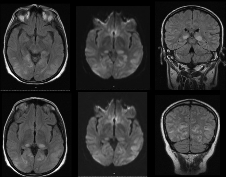
Neurological complications of H1N1 infections are mostly found in children, but rare cases of acute encephalopathy and post-infectious encephalitis such as acute disseminated encephalomyelitis (ADEM) have been described in adults. We report a case of an adult presenting with a progressive and severe encephalopathy that developed after H1N1 respiratory infection resolution. Cerebrospinal fluid (CSF) analysis was normal, including negative PCR for herpes simplex virus, H1N1, influenza B and JC virus, and absent oligoclonal IgG bands in CSF and serum. Initial CT scan was normal, but later MRI showed posterior multifocal leucoencephalopathy with pulvinar sign. The delayed neurological findings together with the ancillary investigation, namely the MRI pattern with both grey and white matter involvement, raised the possibility of a post-infectious process, rather than an acute encephalitis. Despite aggressive immunotherapy, the patient experienced severe neurological sequelae. Early recognition of ADEM manifestations by those dealing with H1N1 infection is important as early immunotherapy may improve the prognosis.
2015 BMJ Publishing Group Ltd.
Figures
Similar articles
-
Encephalitis related to a H1N1 vaccination: case report and review of the literature.Clin Neurol Neurosurg. 2014 Sep;124:8-15. doi: 10.1016/j.clineuro.2014.06.003. Epub 2014 Jun 7. Clin Neurol Neurosurg. 2014. PMID: 24996055 Review.
-
Acute disseminated encephalomyelitis associated with Influenza A H1N1 infection.Neurol Sci. 2011 Oct;32(5):907-9. doi: 10.1007/s10072-011-0500-0. Epub 2011 Mar 8. Neurol Sci. 2011. PMID: 21384278
-
Acute disseminated encephalomyelitis following 2009 H1N1 influenza vaccination.Intern Med. 2012;51(14):1931-3. doi: 10.2169/internalmedicine.51.7487. Epub 2012 Jul 15. Intern Med. 2012. PMID: 22821116
-
A case of acute disseminated encephalomyelitis following influenza virus A-H1N1 infection.Minerva Pediatr. 2013 Oct;65(5):565-7. Minerva Pediatr. 2013. PMID: 24056381
-
Neurological complications of pandemic influenza A H1N1 2009 infection: European case series and review.Eur J Pediatr. 2011 Aug;170(8):1007-15. doi: 10.1007/s00431-010-1392-3. Epub 2011 Jan 14. Eur J Pediatr. 2011. PMID: 21234600 Free PMC article. Review.
Cited by
-
Pulvinar Sign, Stroke and Their Relationship with Fabry Disease: A Systematic Review and Metanalysis.Neurol Int. 2022 Jun 1;14(2):497-505. doi: 10.3390/neurolint14020041. Neurol Int. 2022. PMID: 35736622 Free PMC article. Review.
-
Ocular manifestations of emerging viral diseases.Eye (Lond). 2021 Apr;35(4):1117-1139. doi: 10.1038/s41433-020-01376-y. Epub 2021 Jan 29. Eye (Lond). 2021. PMID: 33514902 Free PMC article. Review.
-
Travellers and influenza: risks and prevention.J Travel Med. 2017 Jan 11;24(1):taw078. doi: 10.1093/jtm/taw078. Print 2017 Jan. J Travel Med. 2017. PMID: 28077609 Free PMC article. Review.
-
Prevention and treatment of respiratory viral infections: Presentations on antivirals, traditional therapies and host-directed interventions at the 5th ISIRV Antiviral Group conference.Antiviral Res. 2018 Jan;149:118-142. doi: 10.1016/j.antiviral.2017.11.013. Epub 2017 Nov 21. Antiviral Res. 2018. PMID: 29162476 Free PMC article.
-
The hidden burden of influenza: A review of the extra-pulmonary complications of influenza infection.Influenza Other Respir Viruses. 2017 Sep;11(5):372-393. doi: 10.1111/irv.12470. Influenza Other Respir Viruses. 2017. PMID: 28745014 Free PMC article. Review.
References
Publication types
MeSH terms
LinkOut - more resources
Full Text Sources
Other Literature Sources
Medical