. 2015 Oct 15;29(20):2123-39.
doi: 10.1101/gad.267278.115.

JMJD1C is required for the survival of acute myeloid leukemia by functioning as a coactivator for key transcription factors

Affiliations

JMJD1C is required for the survival of acute myeloid leukemia by functioning as a coactivator for key transcription factors

Mo Chen et al. Genes Dev. .

Abstract

RUNX1-RUNX1T1 (formerly AML1-ETO), a transcription factor generated by the t(8;21) translocation in acute myeloid leukemia (AML), dictates a leukemic program by increasing self-renewal and inhibiting differentiation. Here we demonstrate that the histone demethylase JMJD1C functions as a coactivator for RUNX1-RUNX1T1 and is required for its transcriptional program. JMJD1C is directly recruited by RUNX1-RUNX1T1 to its target genes and regulates their expression by maintaining low H3K9 dimethyl (H3K9me2) levels. Analyses in JMJD1C knockout mice also establish a JMJD1C requirement for RUNX1-RUNX1T1's ability to increase proliferation. We also show a critical role for JMJD1C in the survival of multiple human AML cell lines, suggesting that it is required for leukemic programs in different AML cell types through its association with key transcription factors.

Keywords: AML1-ETO; HEB; LYL1; acute myeloid leukemia; histone demethylase; transcription regulation.


Figures

Figure 1.
Figure 1.
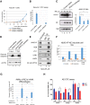
JMJD1C interacts with AETFC in vivo and in vitro. (A, left panel) SDS-PAGE and Commassie staining of HF-AE and associated proteins isolated from Kasumi-1 NE. Immunoprecipitation was performed using NE from Kasumi-1 cells either without (lane 2) or with (lane 3) HF-AE expression. (Lane 1) Protein markers. (Right panel), Identified proteins from this purification. (B) Coimmunoprecipitation and immunoblot confirmation of AE-associated proteins. HF-AE was immunoprecipitated with M2 agarose, and associated proteins were blotted with antibodies to proteins indicated at the left. (C) Immunoprecipitation of JMJD1C to confirm AETFC association. Bound proteins were detected with antibodies shown at the left. (D) Direct interaction between High Five cell-purified f-JMJD1C and HA-AE proteins. Purified HA-AE was immobilized on anti-HA agarose and then incubated with purified f-JMJD1C. Samples were washed with buffers containing increasing amounts of KCl. (E) Immunoprecipitation of HEB to confirm JMJD1C association. Bound proteins were detected with antibodies to proteins shown at the left. (F) JMJD1C interacts more strongly with AETFC than with individual components of the complex. (Top) f-JMJD1C was incubated with the indicated bait proteins immobilized on anti-HA agarose beads. Individual proteins detected by immunoblot are indicated at the left of each blot. Anti-HA antibody was used to detect AE and LMO2, anti-Flag antibody was used to detect JMJD1C, and anti-HEB antibody was used to detect HEB. (G) Direct interaction between f-JMJD1C and E proteins. (Top) Purified E proteins were incubated with f-JMJD1C, and anti-Flag immunoprecipitates were analyzed by immunoblot with antibodies to the proteins indicated at the left.
Figure 2.
Figure 2.
JMJD1C is required for Kasumi-1 cell growth and inhibition of differentiation by AE. (A,B) Assessment of proliferation (A) or colony formation ability (B) of Kasumi-1 cells treated with either a control shRNA lentivirus or two separate JMJD1C shRNA lentiviruses. Error bars represent standard deviation (SD). Data in this figure are represented as mean ± SD. (C) Examination of JMJD1C protein (left) and RNA (right) levels upon shRNA knockdown. (*) P < 0.05. (D) Immunoblot showing increased cleaved caspase 3 upon JMJD1C knockdown. Actin blot served as a loading control. (E) Immunoprecipitation of HF-AE in HL60-HF-AE cells to confirm assembly of intact AETFC complex and JMJD1C association. Bound proteins were detected with antibodies to the proteins shown on the left. (F) Quantification of CD11b-positive cell percentage from flow cytometry experiments indicating AE's ability to inhibit CD11b expression under different differentiation conditions in HL60-HF-AE cells. (G) Bar graph measuring the change of CD11b+ percentage when AE is induced in HL60-HF-AE cells with (lanes 3,4) or without (lanes 1,2) JMJD1C shRNA63. Quantification of three biological replicates of experiments is shown. (*) P < 0.05. (H) Colony counts of AE transformed LIN Jmjd1cf/f bone marrow (BM) cells after transduction of CRE or MIT control viruses in methylcellulose. Results shown are from one wild-type and three f/f mice. After each plating, colony numbers were counted per 10,000 cells plated. (*) P < 0.05.
Figure 3.
Figure 3.
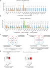
AE directly recruits its cofactor, JMJD1C, to target genes. (AF) ChIP-seq analyses of AE (green) and JMJD1C (red) on known AE-activated target genes LMO2, ID1, EGR1, and CDKN1A and newly identified target genes PADI3 and NOG. Track names are indicated at the left. Gene names are shown below each snapshot. Black bars indicate ChIP-PCR amplicons used in the quantitative PCR (qPCR) analyses in the following figure. (G) Venn diagram depicting numbers of gene promoters (1-kb distance flanking transcription start sites [TSSs]) bound by JMJD1C alone (5848), by AE alone (4210), or jointly by both (2779). P-values assessing significance of the numbers of co-occupied genes are indicated above. (H) Heat map of ChIP-seq reads for JMJD1C and AE rank-ordered from high to low JMJD1C occupancy centered on a ±1-kb window around the TSSs of all genes. Color density reflects read enrichment; white indicates no enrichment. (I) Transcription factor-binding motifs enriched at JMJD1C-binding regions relative to genomic background, with associated P-values indicated at the right of each motif. (J) ChIP-qPCR analyses of JMJD1C occupancy on target genes following control shRNA or AE shRNA treatment. Amplicon positions for peaks on LMO2 (LMO2-p1 and LMO2-pro), ID1 (ID1p1, ID1p2, and ID1p3), CDKN1A (p21-20), and NOG (NOG-pro) genes are schematically indicated as black bars in A, B, D, and F. ChIP amplicons for HBB (HBB-pro), MYC (MYCp1), and SPI1 (PU.1-enh and PU.1-pro) are indicated in Supplemental Figure S4E–G. Data in this figure are represented as mean ± SD. (*) P < 0.05. (K) ChIP-qPCR analyses of AE occupancy using either ETO antibody or HA antibody on target genes CDKN1A (p21) or ID1 in HL60-HF-AE cells. Blue bars and yellow bars represent ChIP assays performed in HL60-HF-AE cells without Dox induction, whereas orange and gray bars represent ChIP assays performed in Dox-induced HL60-HF-AE cells. (*) P < 0.05. (L) ChIP-qPCR analyses of JMJD1C occupancy on target genes upon AE induction in HL60-HF-AE cells. (*) P < 0.05.
Figure 4.
Figure 4.
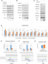
JMJD1C is required for expression of AE target genes. (A,B) RT-qPCR analyses of RNA levels in Kasumi-1 cells treated with either control shRNA, AE shRNA (A), or two separate JMJD1C shRNAs (B). Control shRNA-treated samples were arbitrarily set as 1. Data in this figure are represented as mean ± SD. (*) P < 0.05. (C,D) Venn diagrams showing the numbers of genes differentially expressed with up-regulation (red) or down-regulation (blue) in Kasumi-1 cells treated with JMJD1C shRNA63 (C) or JMJD1C shRNA95 (D) and either control shRNA or AE shRNA. The number of consistently regulated genes is significantly greater (P = 3.5 × 10⁻³¹ or P = 1.8 × 10−56, χ2 test) than the number of oppositely regulated ones. (E) Comparison of RNA expression levels [log(FPKM)] of control shRNA (Control) and AE shRNA-treated (AE) Kasumi-1 cells. (Left panel) Significantly down-regulated genes by JMJD1C knockdown with q-value ≤ 0.05. (Right panel) Significantly up-regulated genes by JMJD1C knockdown with q-value < 0.05. Control and AE at the bottom of each panel indicate the shRNAs used. (***) Significance.
Figure 5.
Figure 5.
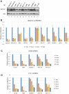
JMJD1C regulates AE target genes by maintaining low H3K9me2 levels. (A) In vitro demethylation assays with purified f-JMJD1C (lane 2) and f-JMJD1A (lane 3) on histones. Antibodies are shown at the left. (B) In vitro demethylation assay with increasing amount of f-JMJD1C on polynucleosomes. (C) In vitro demethylation assay with 293T NEs with (lane 2) or without (lane 1) purified f-JMJD1C. Antibodies are shown at the left. LSD1 is used as loading control for NEs. (D) ChIP-qPCR analyses of H3K9me2 levels on AE target genes in Kasumi-1 cells treated with either control shRNA (blue), AE shRNA (orange), or JMJD1C shRNA63 (gray). ChIP-PCR amplicons are described in Figure 3. Data in this figure are represented as mean ± SD. (*) P < 0.05. (EG, top) ChIP-qPCR analyses of H3K9me2 levels in HL60-HF-AE cells either without (blue) or with (orange) Dox induction on target genes CDKN1A (p21) and ID1 and on nontarget gene HBB. (Bottom) ChIP-seq analyses of AE occupancy in Kasumi-1 cells (green) using anti-ETO antibody or in HL60-HF-AE cells (blue) using anti-HA antibody. ChIP-PCR amplicons are indicated with black bars below the ChIP-seq tracks. The inactive HBB gene is not occupied by either AE or JMJD1C and served as a negative control for H3K9me2 level change. (*) P < 0.05.
Figure 6.
Figure 6.
JMJD1C and HEB/LYL1 are essential for survival of a great variety of AML cells. (A) Immunoblot of JMJD1C protein levels in different leukemic cell NEs. TBP served as a loading control. (BD) Relative proliferation of human leukemia cell lines treated with JMJD1C shRNA63 (B), HEB shRNA (C), or LYL1 shRNA (D). Proliferation was measured by cell viability from 1 d after puromycin selection. Cell numbers obtained from shRNA-treated cells were set to 1. Data in these figures are represented as mean ± SD.
Figure 7.
Figure 7.
HEB/LYL1 are important for JMJD1C recruitment in AMLs. (A) Immunoprecipitation of JMJD1C from different AML cells (indicated below each panel) to confirm HEB and LYL1 association. Bound proteins were detected with antibodies to proteins shown at the left. (B) Heat maps of ChIP-seq reads for JMJD1C, LYL, and AE (only for Kasumi-1 cells) rank-ordered from high to low LYL1 occupancy centered on a ±1-kb window around the peaks of all binding regions of LYL1. Color density reflects read enrichment; white indicates no enrichment. (C,D) HEB and LYL1 are important for JMJD1C occupancy on target genes in Kasumi-1 (C) and NB4 (D) cells. ChIP-qPCR analyses of JMJD1C occupancy on target genes following control shRNA, HEB shRNA, or LYL1 shRNA treatment. Data are represented as mean ± SD. (*) P < 0.05.

Similar articles

Cited by

References

    1. Ben-Ami O, Friedman D, Leshkowitz D, Goldenberg D, Orlovsky K, Pencovich N, Lotem J, Tanay A, Groner Y. 2013. Addiction of t(8;21) and inv(16) acute myeloid leukemia to native RUNX1. Cell Rep 4: 1131–1143. - PubMed
    1. Bernt KM, Zhu N, Sinha AU, Vempati S, Faber J, Krivtsov AV, Feng Z, Punt N, Daigle A, Bullinger L, et al. 2011. MLL-rearranged leukemia is dependent on aberrant H3K79 methylation by DOT1L. Cancer Cell 20: 66–78. - PMC - PubMed
    1. Black JC, Van Rechem C, Whetstine JR. 2012. Histone lysine methylation dynamics: establishment, regulation, and biological impact. Mol Cell 48: 491–507. - PMC - PubMed
    1. Brauchle M, Yao Z, Arora R, Thigale S, Clay I, Inverardi B, Fletcher J, Taslimi P, Acker MG, Gerrits B, et al. 2013. Protein complex interactor analysis and differential activity of KDM3 subfamily members towards H3K9 methylation. PLoS One 8: e60549. - PMC - PubMed
    1. Cabezas-Wallscheid N, Eichwald V, de Graaf J, Lower M, Lehr HA, Kreft A, Eshkind L, Hildebrandt A, Abassi Y, Heck R, et al. 2013. Instruction of haematopoietic lineage choices, evolution of transcriptional landscapes and cancer stem cell hierarchies derived from an AML1-ETO mouse model. EMBO Mol Med 5: 1804–1820. - PMC - PubMed

Publication types

MeSH terms

Substances

Associated data