. 2015 Oct 15;29(20):2140-52.
doi: 10.1101/gad.267997.115.

Constitutive BAK activation as a determinant of drug sensitivity in malignant lymphohematopoietic cells

Affiliations

Constitutive BAK activation as a determinant of drug sensitivity in malignant lymphohematopoietic cells

Haiming Dai et al. Genes Dev. .

Abstract

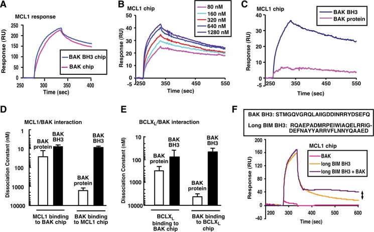
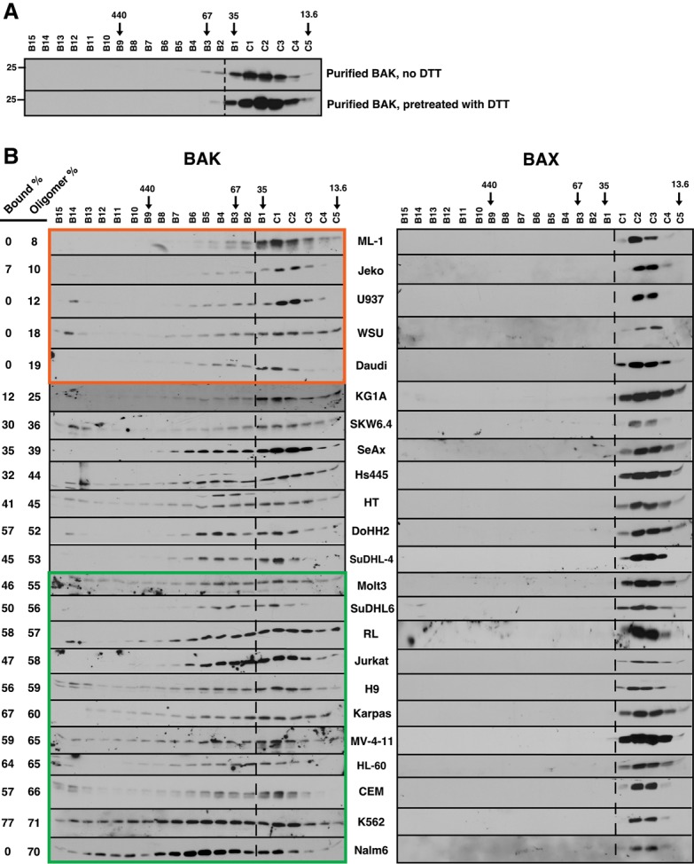
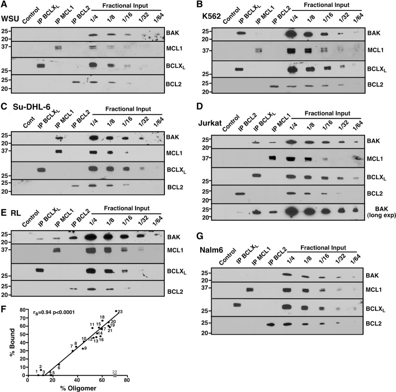
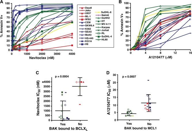
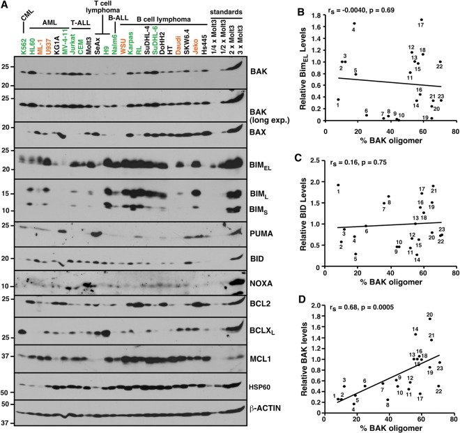
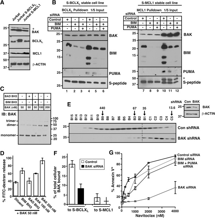
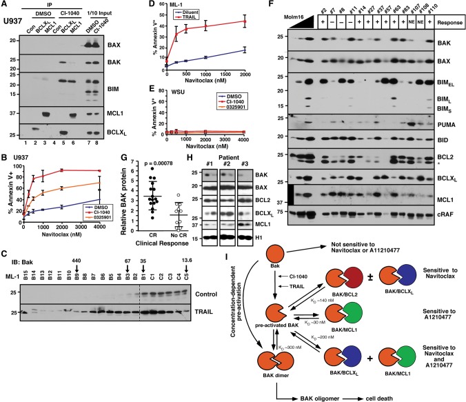
Mitochondrial outer membrane permeabilization (MOMP), a key step in the intrinsic apoptotic pathway, is incompletely understood. Current models emphasize the role of BH3-only BCL2 family members in BAX and BAK activation. Here we demonstrate concentration-dependent BAK autoactivation under cell-free conditions and provide evidence that this autoactivation plays a key role in regulating the intrinsic apoptotic pathway in intact cells. In particular, we show that up to 80% of BAK (but not BAX) in lymphohematopoietic cell lines is oligomerized and bound to anti-apoptotic BCL2 family members in the absence of exogenous death stimuli. The extent of this constitutive BAK oligomerization is diminished by BAK knockdown and unaffected by BIM or PUMA down-regulation. Further analysis indicates that sensitivity of cells to BH3 mimetics reflects the identity of the anti-apoptotic proteins to which BAK is constitutively bound, with extensive BCLXL•BAK complexes predicting navitoclax sensitivity, and extensive MCL1•BAK complexes predicting A1210477 sensitivity. Moreover, high BAK expression correlates with sensitivity of clinical acute myelogenous leukemia to chemotherapy, whereas low BAK levels correlate with resistance and relapse. Collectively, these results inform current understanding of MOMP and provide new insight into the ability of BH3 mimetics to induce apoptosis without directly activating BAX or BAK.

Keywords: BAK; BH3 mimetic; apoptosis.


Figures

Figure 1.
Figure 1.
MCL1 and BCLXL preferentially bind activated BAK. (A) SPR of 300 nM MCL1ΔTM binding to immobilized BAKΔTM or BAK BH3 peptide. (B) SPR (relative units) of different concentrations of BAK BH3 peptide binding to immobilized MCL1ΔTM. (C) Direct comparison of 320 nM BAKΔTM and 320 nM BAK BH3 peptide binding to immobilized MCL1ΔTM. (D,E) Based on the SPR assays, the affinities of MCL1ΔTM (D) and BCLXLΔTM (E) for BAKΔTM and BAK BH3 peptide were calculated. Error bars indicate ±sd of three independent experiments using different chips and different protein batches. (F) SPR of MCL1ΔTM binding to 500 nM BAKΔTM, 500 nM BIM BH3, or 500 nM BAKΔTM + 500 nM BIM BH3. The double arrow indicates increased resonance due to BAK activation. The box at the top of F indicates peptides used in this figure.
Figure 2.
Figure 2.
BAK, but not BAX, is constitutively oligomerized in malignant lymphohematopoietic cell lines. (A) Purified BAKΔTM (20 µM) without or with 5 mM DTT was incubated in isotonic buffer containing 1% CHAPS for 1 h and subjected to size exclusion chromatography. (B) Lysates prepared from the indicated log-phase cells in 1% CHAPS were subjected to size exclusion chromatography and blotted with antibodies to BAK (left) or BAX (right). Numbers at the left indicate the sum of the total percentage of BAK that is detected as bound to BCLXL + MCL1 + BCL2 in pull-down assays (“Bound %”) (Fig. 3; Supplemental Fig S5), calculated as described (Dai et al. 2009), and on FPLC assays (running to the left of the dashed line) (“Oligomer %”), calculated as indicated in Supplemental Figure S2. Arrows indicate size markers in kilodaltons. Orange and green boxes indicate cell lines with low (<20%) and high (>55%) constitutive BAK oligomerization, respectively.
Figure 3.
Figure 3.
BAK is constitutively bound to various anti-apoptotic BCL2 family members. (AE,G) CHAPS lysates of WSU (A), K562 (B), Su-DHL-6 (C), Jurkat (D), RL (E), and Nalm6 (G) cells were immunoprecipitated (IP) with antibodies to BCLXL, MCL1, or BCL2, and compared with serial dilutions of the input. Pull-downs with normal rabbit IgG served as a control. (F) The relative amount of BAK bound to BCL2 + BCLXL + MCL1 (% Bound) compared with the relative amount of oligomerized BAK (% Oligomerized), calculated as indicated in Supplemental Figure S2. Cell lines used here and in Figure 5 and Supplemental Figure S8 are ML-1 (1), Jeko (2), U937 (3), WSU (4), Daudi (5), KG1A (6), SKW6.4 (7), SeAx (8), Hs445 (9), HT (10), DoHH2 (11), SuDHL-4 (12), Molt3 (13), SuDHL-6 (14), RL (15), Jurkat (16), H9 (17), Karpas422 (18), MV-4-11 (19), HL-60 (20), CEM (21), Nalm6 (22), and K562 (23). As indicated in the text, Nalm6 was excluded from subsequent calculations.
Figure 4.
Figure 4.
Sensitivities of cell lines to navitoclax and A1210477 correlate with BAK binding status. (A,B) After the cell lines were treated with navitoclax (A) or A1210477 (B), the percentages of Annexin V-positive cells were assessed by flow cytometry. Graphs show the average of three independent experiments. Symbols and line colors represent different BAK binding statuses. (Red) No binding; (blue) BAK bound to BCLXL only; (yellow) BAK bound to MCL1 only; (purple) unknown binding partner; (green) BAK bound to multiple partners. (C,D) Concentrations of navitoclax (C) or A1210477 (D) required to induce apoptosis in 50% of cells (IC50) grouped according to BAK binding status as determined in Figure 3 and Supplemental Figure S5. Symbols for each cell line correspond to the key between A and B. Heavy horizontal lines represent the mean, and error bars indicate the SD of the indicated groups of lines.
Figure 5.
Figure 5.
Constitutive BAK oligomerization correlates with relative BAK content. (A) Whole-cell lysates, along with different concentrations of Molt3, were blotted with the indicated antibodies. Orange and green letters indicate the cell lines in the orange and green boxes, respectively, in Figure 2. (BD) The percentage of BAK that is constitutively oligomerized (% BAK oligomer), calculated as shown in Supplemental Figure S2, versus the amount of BIMEL (B), BID (C), or BAK (D) in each line relative to an equal amount of Molt3 whole-cell lysate, determined as described in the Materials and Methods.
Figure 6.
Figure 6.
Constitutive BAK oligomerization is inhibited by BAK knockdown. (A) Jurkat sublines stably expressing BCLXL or MCL1 tagged at the N terminus with S peptide (S-BCLXL or S-MCL1). (B) Forty-eight hours after cells in A were transfected with the indicated siRNAs, proteins were pulled down on S peptide-agarose and, along with aliquots of the lysates (“input”), subjected to immunoblotting. (C) After the indicated concentrations of BAKΔTM without or with BH3 peptides were incubated with liposomes for 30 min at 37°C, BAK oligomers were cross-linked with bismaleimidohexane and detected by immunoblotting. Note the increasing dimer and trimer with increasing BAK concentrations. (D) Release of encapsulated FITC-dextran from liposomes during a 1-h incubation as illustrated in Supplemental Figure S11A. (E) Forty-eight hours after Jurkat cells were transfected with BAK shRNA, CHAPS lysates were fractioned by FPLC and blotted with BAK antibody. (Inset) Western blot of whole-cell lysates. (F) Forty-eight hours after the cells shown in A were transfected with BAK siRNA or nontargeting control, the percentages of BAK bound to S-BCLXL and S-MCL1 were calculated from data shown in Supplemental Figure S12, B and C. (G) Twenty-four hours after Jurkat cells were transfected with the indicated siRNAs, cells were treated with navitoclax for another 24 h and stained with Annexin V. Error bars indicate ±sd of three independent assays (D,G) or a range of two independent assays (F). (*) Nonspecific band in A and B.
Figure 7.
Figure 7.
Drug-induced BAK oligomerization sensitizes to navitoclax. (A) After U937 cells were treated with 10 µM CI-1040 for 24 h, BCLXL and MCL1 were immunoprecipitated and compared with 1/10 of input. Normal rabbit IgG antibody served as an immunoprecipitation control. (B,D,E) After U937 (B), ML-1 (D), or WSU (E) cells were treated with the indicated drugs for 24 h, apoptosis was detected by Annexin V staining. (C) Twenty-four hours after ML-1 cells were treated with 12.5 ng/mL TRAIL, CHAPS lysates were subjected to size exclusion FPLC and blotted for BAK. (F) Clinical acute myelogenous leukemia (AML) samples harvested at initial diagnosis together with different concentrations of Molm16 cell lines as control were blotted with the indicated antibodies. (+) Attainment of complete remission (CR); (−) no CR; (NE) not evaluable due to death before remission status determined. (G) Relative BAK expression, calculated as BAKx ÷ RAFx, where BAKx and RAFx are the signals for BAK and the housekeeping protein cRAF in 5 × 10⁵ leukemia cells from patient X relative to the serial dilution of Molm16 cells (see F), compared in two groups according to patient outcome. (H) Sequential AML samples harvested at diagnosis and relapse were blotted with the indicated antibodies. (I) Model of concentration-dependent BAK activation described in the Discussion. Equilibrium dissociation constants (KDs) for various complexes were derived from SPR experiments shown in Figure 1 and Supplemental Figure S15 or as previously published (Dai et al. 2009).

Similar articles

Cited by

References

    1. Antonsson B, Montessuit S, Sanchez B, Martinou JC. 2001. Bax is present as a high molecular weight oligomer/complex in the mitochondrial membrane of apoptotic cells. J Biol Chem 276: 11615–11623. - PubMed
    1. Brouwer JM, Westphal D, Dewson G, Robin AY, Uren RT, Bartolo R, Thompson GV, Colman PM, Kluck RM, Czabotar PE. 2014. Bak core and latch domains separate during activation, and freed core domains form symmetric homodimers. Mol Cell 55: 938–946. - PubMed
    1. Certo M, Moore Vdel G, Nishino M, Wei G, Korsmeyer S, Armstrong SA, Letai A. 2006. Mitochondria primed by death signals determine cellular addiction to antiapoptotic BCL-2 family members. Cancer Cell 9: 351–365. - PubMed
    1. Cheng EH, Sheiko TV, Fisher JK, Craigen WJ, Korsmeyer SJ. 2003. VDAC2 inhibits BAK activation and mitochondrial apoptosis. Science 301: 513–517. - PubMed
    1. Chipuk JE, Green DR. 2008. How do BCL-2 proteins induce mitochondrial outer membrane permeabilization? Trends Cell Biol 18: 157–164. - PMC - PubMed

Publication types

MeSH terms