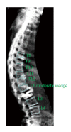
Vertebral fracture assessment: Current research status and application in patients with kyphoplasty
- PMID: 26495245
- PMCID: PMC4610910
- DOI: 10.5312/wjo.v6.i9.680
Vertebral fracture assessment: Current research status and application in patients with kyphoplasty
Abstract
Imaging of the spine is of paramount importance for the recognition of osteoporotic vertebral fractures (VFs), and standard radiography (SR) of the spine is the suggested diagnostic method but is not routinely used because of the cost and radiation exposure considerations. VF assessment (VFA) is an efficient, low radiation method for identifying VFs at the time of bone mineral density (BMD) measurement. Prediction models used to indicate the need for VFA may have little predictive power in subspecialty referral populations such as rheumatologic patients or patients who underwent kyphoplasty. Rheumatologic patients are frequently at increased risk for VFs, and VFA should be performed on an individual basis, also taking in account the guidelines for the general population. Kyphoplasty is a new minimal invasive procedure for the treatment of VFs and is being performed with increasing frequency. Following kyphoplasty, there may be a risk of new VFs in adjacent vertebrae. The assessment and follow-up of patients who underwent kyphoplasty requires repetitive X-ray imaging with the known limitations of SR. Thus, VFA may facilitate the evaluation of VFs in these patients because most of the kyphoplasty patients would fulfill the criteria. In a pilot study, we measured the BMD and performed VFA in 28 patients treated with kyphoplasty. Ratios of anterior to posterior (A/P) and middle to posterior (M/P) height were measured, and Genant's method was used to classify vertebrae accordingly. Intraobserver and interobserver reliability for A/P, M/P and the Genant's method were determined. Only 1 patient did not meet the criteria for VFA. Of the 364 available vertebrae, 295 could be analyzed. Most missing data (concerning 69 vertebrae) occurred in the upper thoracic region. Three of the 69 non-eligible vertebrae were lumbar vertebrae with cement leakage from the kyphoplasty procedure. In our hands, VFA was highly reproducible, demonstrating very good agreement in terms of intraobserver and interobserver reliability. Agreement was very good on the vertebral level, "vertebrae with kyphoplasty" level and "2 above and 1 below the kyphoplasty vertebrae" level. The application of Genant's method to these patients also resulted in perfect agreement. We believe that the potential value of VFA in patients treated with kyphoplasty requires further evaluation, particularly comparing VFA with SR and performing a longitudinal follow-up. More research will help to adopt care processes that determine which patients require VFA and how often VFA should be performed, while also considering the impact of this technique on the cost of healthcare organizations.
Keywords: Current research; Guideline; Kyphoplasty; Vertebral fracture assessment.
Figures


References
-
- Cummings SR, Melton LJ. Epidemiology and outcomes of osteoporotic fractures. Lancet. 2002;359:1761–1767. - PubMed
-
- Fink HA, Milavetz DL, Palermo L, Nevitt MC, Cauley JA, Genant HK, Black DM, Ensrud KE. What proportion of incident radiographic vertebral deformities is clinically diagnosed and vice versa? J Bone Miner Res. 2005;20:1216–1222. - PubMed
-
- Melton LJ, Lane AW, Cooper C, Eastell R, O’Fallon WM, Riggs BL. Prevalence and incidence of vertebral deformities. Osteoporos Int. 1993;3:113–119. - PubMed
-
- Bouxsein ML, Melton LJ, Riggs BL, Muller J, Atkinson EJ, Oberg AL, Robb RA, Camp JJ, Rouleau PA, McCollough CH, et al. Age- and sex-specific differences in the factor of risk for vertebral fracture: a population-based study using QCT. J Bone Miner Res. 2006;21:1475–1482. - PubMed
-
- Jinbayashi H, Aoyagi K, Ross PD, Ito M, Shindo H, Takemoto T. Prevalence of vertebral deformity and its associations with physical impairment among Japanese women: The Hizen-Oshima Study. Osteoporos Int. 2002;13:723–730. - PubMed
LinkOut - more resources
Full Text Sources
Other Literature Sources
Research Materials

