. 2015 Oct 23;11(10):e1005609.
doi: 10.1371/journal.pgen.1005609. eCollection 2015 Oct.

Virus Satellites Drive Viral Evolution and Ecology

Affiliations

Virus Satellites Drive Viral Evolution and Ecology

Belén Frígols et al. PLoS Genet. .

Abstract

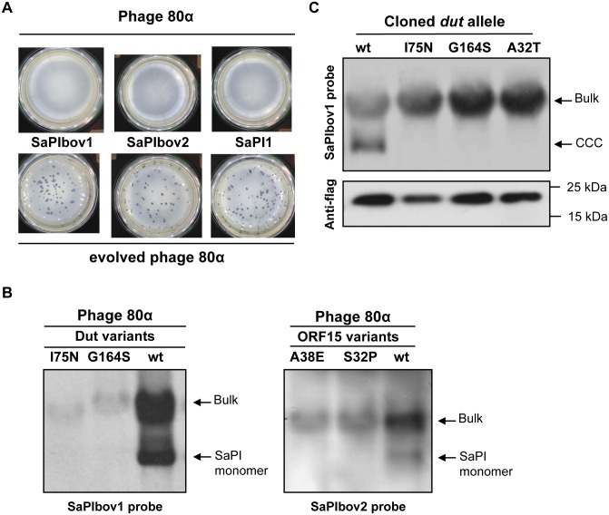
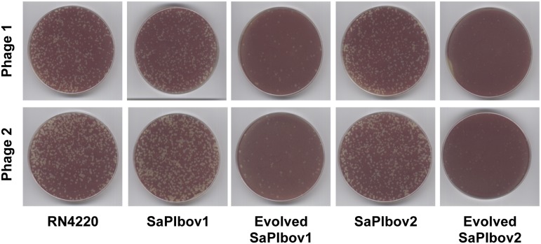
Virus satellites are widespread subcellular entities, present both in eukaryotic and in prokaryotic cells. Their modus vivendi involves parasitism of the life cycle of their inducing helper viruses, which assures their transmission to a new host. However, the evolutionary and ecological implications of satellites on helper viruses remain unclear. Here, using staphylococcal pathogenicity islands (SaPIs) as a model of virus satellites, we experimentally show that helper viruses rapidly evolve resistance to their virus satellites, preventing SaPI proliferation, and SaPIs in turn can readily evolve to overcome phage resistance. Genomic analyses of both these experimentally evolved strains as well as naturally occurring bacteriophages suggest that the SaPIs drive the coexistence of multiple alleles of the phage-coded SaPI inducing genes, as well as sometimes selecting for the absence of the SaPI depressing genes. We report similar (accidental) evolution of resistance to SaPIs in laboratory phages used for Staphylococcus aureus typing and also obtain the same qualitative results in both experimental evolution and phylogenetic studies of Enterococcus faecalis phages and their satellites viruses. In summary, our results suggest that helper and satellite viruses undergo rapid coevolution, which is likely to play a key role in the evolution and ecology of the viruses as well as their prokaryotic hosts.


Conflict of interest statement

The authors have declared that no competing interests exist.

Figures

Fig 1
Fig 1. SaPI interference in evolved phages.
(A) SaPI interference with phage reproduction. Approximately 108 bacteria were infected with 100 p.f.u. of phage 80α (upper panel) or an evolved 80α derivative carrying mutation in all three SaPI inducers (lower panel), plated on phage bottom agar, and incubated 24 h at 32°C. (B) Induction of SaPIbov1 (left) or SaPIbov2 (right) by evolved 80α phages carrying mutations in the dut or ORF15 genes, respectively. Samples from the different lysogenic strains were isolated 60 min after induction with mitomycin C, separated on agarose and blotted with a SaPIbov1- or SaPIbov2-specific probe. Upper band is ‘bulk’ DNA, including chromosomal, phage and replicating SaPI; lower band is SaPI linear monomers released from phage heads. (C) SaPIbov1 excision and replication after induction of cloned dut genes from different evolved phages. A non-lysogenic derivative of strain RN4220 carrying SaPIbov1 was complemented with plasmids expressing 3xFlag-tagged Dut proteins. One millilitre of each culture (optical density (OD)540nm = 0.3) was collected and used to prepare standard minilysates, which were resolved on a 0.7% agarose gel, Southern blotted and probed for SaPIbov1 DNA. In these experiments, because no helper phage is present, the excised SaPI DNA appears as covalently closed circular molecules (CCC) rather than the linear monomers that are seen following helper-phage-mediated induction and packaging. The upper panel is a Southern blot probed for SaPIbov1 DNA; the lower panel is a western blot probed with antibody (Sigma) to the Flag tag carried by the proteins.
Fig 2
Fig 2. Coevolved SaPIs block phage reproduction.
Plates carrying the SaPI-negative RN4220 strain, RN4220 derivatives carrying the original SaPIbov1 or SaPIbov2 islands, or RN4220 derivatives carrying evolved SaPIbov1 or SaPIbov2 islands were infected (<700 p.f.u. per plate) with two different evolved phage 80α. Genotype of the SaPIbov1 evolved island: A deleted from position 14119; genotype of the SaPIbov2 evolved island: deletion affecting residues from N122 to K168. Phage 1: 80α Dut I75N, ORF15 Q3*, Δsri; Phage 2: Dut S63I, ORF15 A38E, sri G 10983 A.
Fig 3
Fig 3. SaPI-driven phage evolution occurs in vivo.
(A) Partial genetic maps of ϕ55–2 and ϕ55–3 (GenBank accession numbers KR709302 and KR709303, respectively). Arrows indicate predicted ORFs. Coloured arrows indicate the divergent region found in these phages, which include the SaPI1 inducing genes. (B) Lineup of Sri (SaPI1 inducer) protein sequences from phages ϕ55–2 and ϕ55–3, coloured according to relative sequence conservation at each position. Adapted from lineup generated by PRALINE [41]. The scoring scheme works from 0 for the least conserved alignment position, up to 10 (asterisk) for the most conserved alignment position. (C) Lineup of Dut (SaPIbov1 inducer) protein sequences from phages 80α and ϕSaov3 (left) or from phages ϕ11 and ϕB2 (right), coloured according to relative sequence conservation at each position. Adapted from lineup generated by PRALINE [41]. (D) SaPIbov1 excision and replication after induction of cloned dut genes from the natural mutant phages analysed in (C). A non-lysogenic derivative of strain RN4220 carrying SaPIbov1 was complemented with plasmids expressing 3xFlag-tagged Dut proteins, as indicated in Fig 1. The upper panel is a Southern blot probed for SaPIbov1 DNA; the lower panel is a western blot probed with antibody (Sigma) to the Flag tag carried by the proteins.
Fig 4
Fig 4. The enterococcal EfCIV583 island drives phage evolution.
(A) Nearest neighbour tree of EfsCIV583 inducer proteins generated by MEGA5 [36]. Numbers indicate the bootstrap value. Shaded are the proteins characterised in this study. (B) Lineup of selected EfCIV583 inducer protein sequences from different enterococcal phages and prophages, coloured according to relative sequence conservation at each position. Adapted from lineup generated by PRALINE [41]. Accession numbers EfsCIV583 inducer proteins: EF0309 (AAO80172); Ef11 (YP_003358829); X98 (WP_002381619); VC1B-1 (EPI33180).

Similar articles

Cited by

References

    1. Huang Y-W, Hu C-C, Lin N-S, Hsu Y-H. Mimicry of molecular pretenders: the terminal structures of satellites associated with plant RNA viruses. RNA Biol. 2010;7: 162–171. - PubMed
    1. Nawaz-ul-Rehman MS, Fauquet CM. Evolution of geminiviruses and their satellites. FEBS Lett. 2009;583: 1825–1832. 10.1016/j.febslet.2009.05.045 - DOI - PubMed
    1. Simon AE, Roossinck MJ, Havelda Z. Plant virus satellite and defective interfering RNAs: new paradigms for a new century. Annu Rev Phytopathol. 2004;42: 415–437. - PubMed
    1. Christie GE, Dokland T. Pirates of the Caudovirales. Virology. 2012;434: 210–221. 10.1016/j.virol.2012.10.028 - DOI - PMC - PubMed
    1. Ram G, Chen J, Kumar K, Ross HF, Ubeda C, Damle PK, et al. Staphylococcal pathogenicity island interference with helper phage reproduction is a paradigm of molecular parasitism. Proc Natl Acad Sci USA. 2012;109: 16300–16305. 10.1073/pnas.1204615109 - DOI - PMC - PubMed

Publication types

MeSH terms