

The effect of chemotherapy on the mammographic appearance of breast cancer and correlation with histopathology
- PMID: 26495873
- PMCID: PMC4985959
- DOI: 10.1259/bjr.20150479
The effect of chemotherapy on the mammographic appearance of breast cancer and correlation with histopathology
Abstract
Objective: To document the mammographic changes after neoadjuvant chemotherapy with histopathological correlation, to calculate the accuracy of mammography (MG) in predicting residual tumour size and to measure the interobserver agreement in reading mammograms.
Methods: In 446 consecutive cases, the pre- and post-chemotherapy mammograms were retrospectively evaluated by two blinded observers, and consensus findings were compared with reference standard of surgical specimen. The accuracy of MG in predicting residual tumour size was calculated. Kappa statistics were calculated for measuring the interobserver agreement for reading mammograms. The sensitivity, specificity, positive-predictive value and negative-predictive value for the prediction of residual disease were calculated.
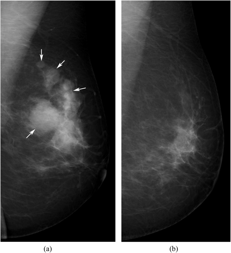
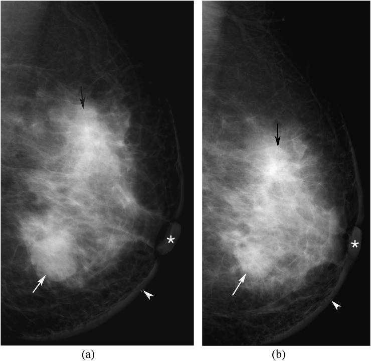
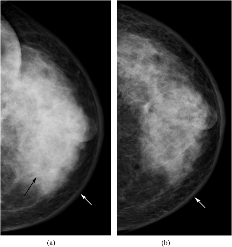
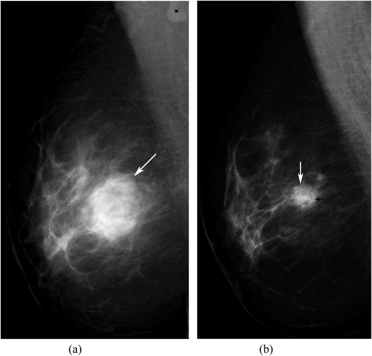
Results: The most common primary abnormalities were mass lesions without and with microcalcifications. After chemotherapy, there was decrease in size of most (95.1%) of the measurable masses, with decrease in the mean tumour size from 4.1 to 2.5 cm. The density of the tumour decreased in 66.6% (241/362) cases with residual disease. There was almost perfect interobserver agreement for describing the primary abnormality in the pre- as well as post-chemotherapy mammograms (k = 0.87 and 0.81, respectively) with substantial agreement for measurement of the mass lesions before and after chemotherapy (k = 0.69 and 0.68, respectively). MG showed accuracy of 60.0%, sensitivity of 94.4%, specificity of 50.0%, positive-predictive value of 91.3% and negative-predictive value of 61.8%.
Conclusion: MG remains a highly sensitive and reproducible investigation for the assessment of residual disease after chemotherapy.
Advances in knowledge: There is substantial interobserver agreement in characterizing and measuring breast tumours on mammograms.
Figures
Similar articles
-
Accuracy of physical examination, ultrasonography, and mammography in predicting residual pathologic tumor size in patients treated with neoadjuvant chemotherapy.Ann Surg. 2006 Feb;243(2):257-64. doi: 10.1097/01.sla.0000197714.14318.6f. Ann Surg. 2006. PMID: 16432360 Free PMC article.
-
Mammography in the follow-up after breast-conserving treatment in cancer of the breast: suitability for mammographic interpretation, validity and interobserver variation.Br J Radiol. 1995 Jul;68(811):754-60. doi: 10.1259/0007-1285-68-811-754. Br J Radiol. 1995. PMID: 7640932
-
Residual Mammographic Microcalcifications and Enhancing Lesions on MRI After Neoadjuvant Systemic Chemotherapy for Locally Advanced Breast Cancer: Correlation with Histopathologic Residual Tumor Size.Ann Surg Oncol. 2016 Apr;23(4):1135-42. doi: 10.1245/s10434-015-4993-2. Epub 2015 Dec 1. Ann Surg Oncol. 2016. PMID: 26628432
-
Accuracy of the combination of mammography and sonography in predicting tumor response in breast cancer patients after neoadjuvant chemotherapy.Ann Surg Oncol. 2006 Nov;13(11):1443-9. doi: 10.1245/s10434-006-9086-9. Epub 2006 Sep 21. Ann Surg Oncol. 2006. PMID: 17028770 Clinical Trial.
-
Meta-analysis of agreement between MRI and pathologic breast tumour size after neoadjuvant chemotherapy.Br J Cancer. 2013 Sep 17;109(6):1528-36. doi: 10.1038/bjc.2013.473. Epub 2013 Aug 20. Br J Cancer. 2013. PMID: 23963140 Free PMC article. Review.
Cited by
-
The role and potential of digital breast tomosynthesis in neoadjuvant systemic therapy evaluation for optimising breast cancer management: a pictorial essay.Br J Radiol. 2025 Apr 1;98(1168):485-495. doi: 10.1093/bjr/tqae252. Br J Radiol. 2025. PMID: 39724185 Free PMC article. Review.
-
Predictive value of ultrasound doppler parameters in neoadjuvant chemotherapy response of breast cancer: Prospective comparison with magnetic resonance and mammography.PLoS One. 2024 Jun 4;19(6):e0302527. doi: 10.1371/journal.pone.0302527. eCollection 2024. PLoS One. 2024. PMID: 38833499 Free PMC article.
-
Construction of Nomograms for Predicting Pathological Complete Response and Tumor Shrinkage Size in Breast Cancer.Cancer Manag Res. 2020 Sep 10;12:8313-8323. doi: 10.2147/CMAR.S270687. eCollection 2020. Cancer Manag Res. 2020. PMID: 32982426 Free PMC article.
-
Imaging findings for response evaluation of ductal carcinoma in situ in breast cancer patients treated with neoadjuvant systemic therapy: a systematic review and meta-analysis.Eur Radiol. 2023 Aug;33(8):5423-5435. doi: 10.1007/s00330-023-09547-7. Epub 2023 Apr 5. Eur Radiol. 2023. PMID: 37020070 Free PMC article.
References
-
- Ferlay J, Soerjomataram I, Ervik M, Dikshit R, Eser S, Mathers C, et al. . GLOBOCAN 2012 V1.0, Cancer Incidence and Mortality Worldwide: IARC Cancer Base No 11. Lyon, France: International Agency for Research on Cancer; 2013 [Updated 9 September 2014; cited 11 April 2015]. Available from: http://globocan.iarc.fr
-
- Calais G, Berger C, Descamps P, Chapet S, Reynaud-Bougnoux A, Body G, et al. . Conservative treatment feasibility with induction chemotherapy, surgery, and radiotherapy for patients with breast carcinoma larger than 3 cm. Cancer 1994; 74: 1283–8. doi: 10.1002/1097-0142(19940815)74:4<1283::AID-CNCR2820740417>3.0.CO;2-S - DOI - PubMed
-
- Booser DJ, Hortobagyi GN. Treatment of locally advanced breast cancer. Semin Oncol 1992; 19: 278–85. - PubMed
MeSH terms
LinkOut - more resources
Full Text Sources
Other Literature Sources
Medical
