. 2016 Mar;216(3):358-75.
doi: 10.1111/apha.12622. Epub 2015 Nov 11.

Deficiency or inhibition of lysophosphatidic acid receptor 1 protects against hyperoxia-induced lung injury in neonatal rats

Affiliations

Deficiency or inhibition of lysophosphatidic acid receptor 1 protects against hyperoxia-induced lung injury in neonatal rats

X Chen et al. Acta Physiol (Oxf). 2016 Mar.

Abstract

Aim: Blocking of lysophosphatidic acid (LPA) receptor (LPAR) 1 may be a novel therapeutic option for bronchopulmonary dysplasia (BPD) by preventing the LPAR1-mediated adverse effects of its ligand (LPA), consisting of lung inflammation, pulmonary arterial hypertension (PAH) and fibrosis.

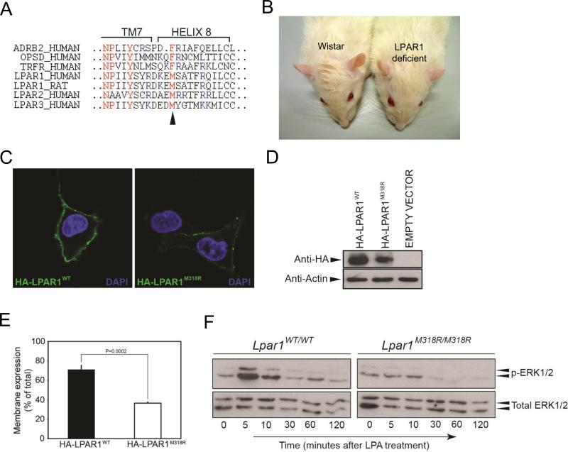
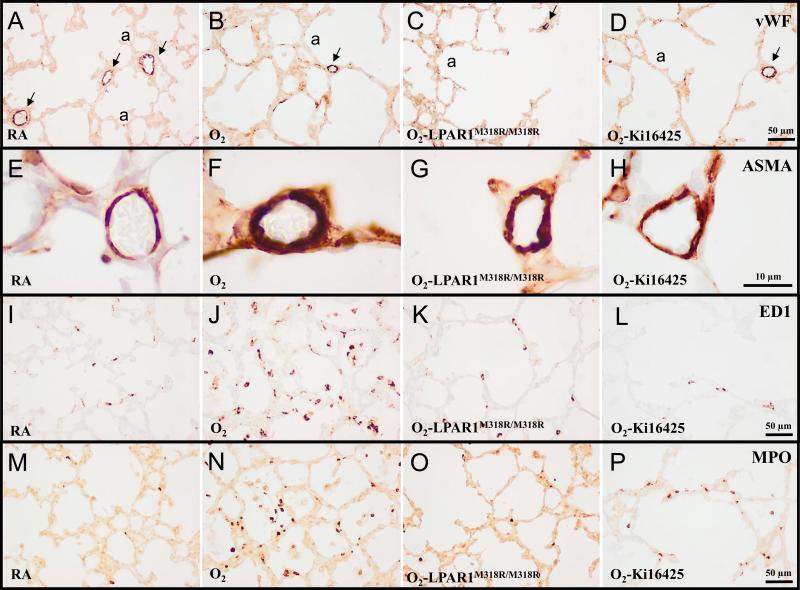
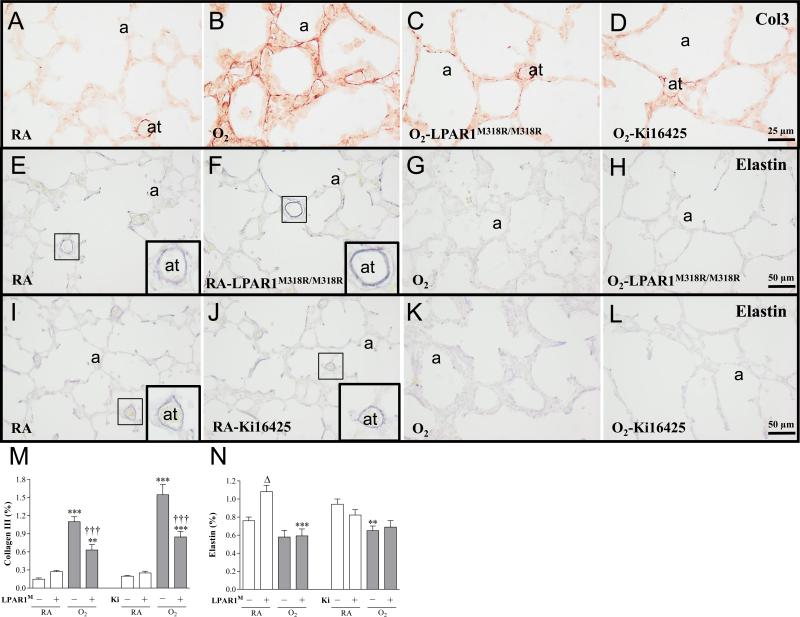
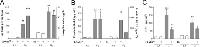
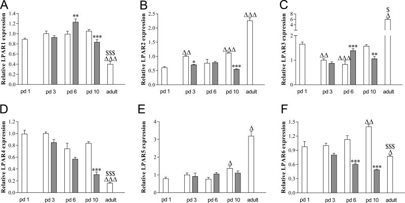
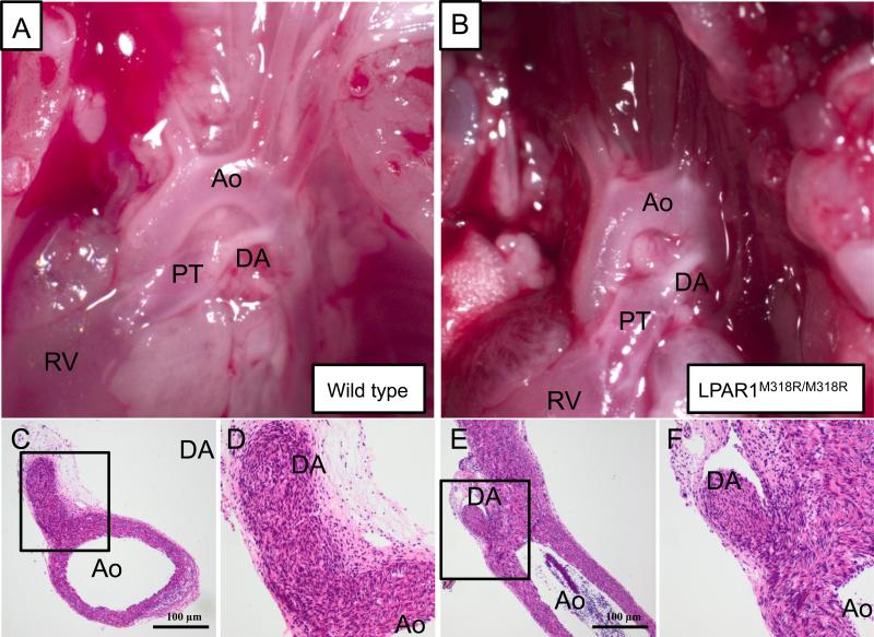
Methods: In Wistar rats with experimental BPD, induced by continuous exposure to 100% oxygen for 10 days, we determined the beneficial effects of LPAR1 deficiency in neonatal rats with a missense mutation in cytoplasmic helix 8 of LPAR1 and of LPAR1 and -3 blocking with Ki16425. Parameters investigated included survival, lung and heart histopathology, fibrin and collagen deposition, vascular leakage and differential mRNA expression in the lungs of key genes involved in LPA signalling and BPD pathogenesis.

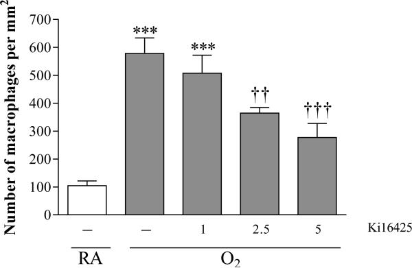
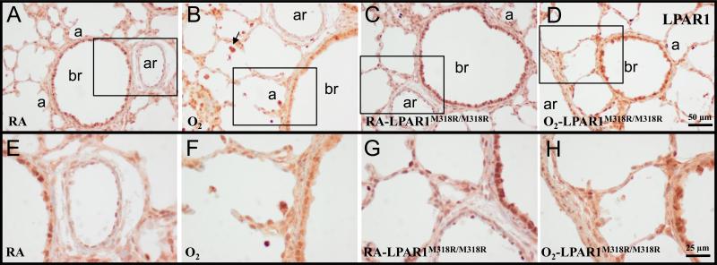
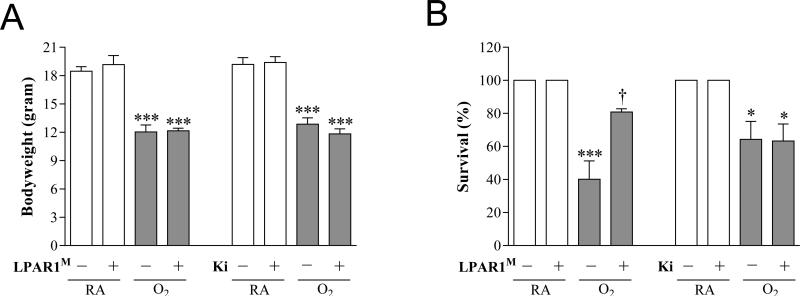
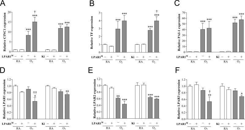
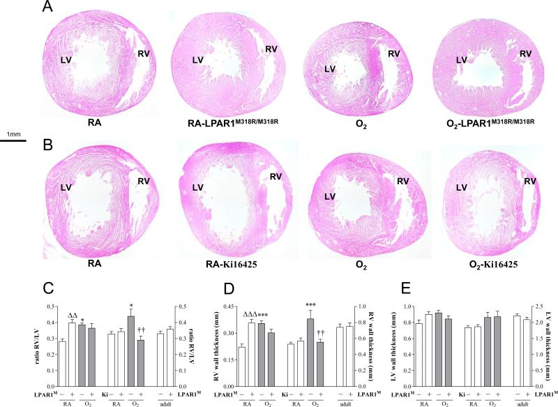
Results: LPAR1-mutant rats were protected against experimental BPD and mortality with reduced alveolar septal thickness, lung inflammation (reduced influx of macrophages and neutrophils, and CINC1 expression) and collagen III deposition. However, LPAR1-mutant rats were not protected against alveolar enlargement, increased medial wall thickness of small arterioles, fibrin deposition and vascular alveolar leakage. Treatment of experimental BPD with Ki16425 confirmed the data observed in LPAR1-mutant rats, but did not reduce the pulmonary influx of neutrophils, CINC1 expression and mortality in rats with experimental BPD. In addition, Ki16425 treatment protected against PAH and right ventricular hypertrophy.

Conclusion: LPAR1 deficiency attenuates pulmonary injury by reducing pulmonary inflammation and fibrosis, thereby reducing mortality, but does not affect alveolar and vascular development and, unlike Ki16425 treatment, does not prevent PAH in neonatal rats with experimental BPD.

Keywords: bronchopulmonary dysplasia; fibrosis; lung inflammation; lysophosphatidic acid receptor; right ventricular hypertrophy.


Figures

Figure 1
Figure 1
Pilot experiment to find the optimal dose of Ki16425 for treatment of experimental BPD by determining the pulmonary influx of ED-1-positive monocytes and macrophages in tissue sections in room air (RA), pups injected daily with DMSO (10%, open bar) and O2-exposed pups (O2) injected daily with 10% DMSO or Ki16425 (shaded bars): 1, 2.5 and 5 mg kg−1 day−1. Data are expressed as mean ± SEM. ***p < 0.001 versus RA control. ††p <0.01 and †††p < 0.001 versus age-matched DMSO-treated O2-exposed control; n=4-8 per group.
Figure 2
Figure 2
Representative lung sections stained for lysophosphatidic acid receptor 1 (LPAR1 or endothelial differentiation gene 2 (EDG2); A-H) of wild type (A, B, E and F) and LPAR1 mutant rat pups (C, D, G and H) kept in room-air (RA; A, C, E and G) or 100% O2 (B, D, F and H) until 10 days of age. Boxed areas in panels A, B, C and D are represented in panels E, F, G and H, respectively. a = alveolus; ar = arteriole; br = bronchus. Arrow in panel B indicates LPAR1-positive inflammatory cells.
Figure 3
Figure 3
Nonsynonymous mutation in the LPAR1 gene results in a hypomorphic phenotype. (A) Alignment of amino acid sequence of the 8th helices of different class A GPCRs. Indicated in red is the NPxxY(x)5,6F motif of which the tyrosine in transmembrane (TM) 7 domain and the phenylalanine in helix 8 form a hydrophobic interaction in the inactive state. In LPAR1-3 the phenylalanine is replaced by a methionine, conserving hydrophobicity. The arrowhead indicates the location of the amino acid change caused by the mutation. Indicated in blue are the basic amino acids that are commonly found in helix 8. (B) LPAR1 mutant rats have a craniofacial malformation with a shortening of the eye to nose tip distance and an increase in the distance between the eyes compared to wild type Wistar rats. (C) Decreased membrane expression of overexpressed LPAR1M318R compared to overexpressed wild-type receptor. Intact serum-starved COS-7 cells expressing N-terminally HA-tagged wild-type or mutant LPAR1 were incubated with an anti-HA antibody. The antibody can only bind if the cell expresses the receptor in the membrane, because only then the HA-tag is localized outside the cell. (D) Western blot analysis of COS-7 cells overexpressing either HA-LPAR1WT or HA-LPAR1M318R. (E) The size of the cell surface expressed receptor pool as percentage of the total receptor pool is decreased in cells expressing LPAR1M318R. Graph indicates the pool of cell surface expressed receptor as a percentage of total HA-tagged expressed receptors measured by cell surface ELISA. (F) LPAR1M318R/M318R rat embryonic fibroblasts (REFs) display a hypomorphic ERK1/2 phosphorylation pattern. Western blot analysis of MAP kinase pathway activation in wild type and mutant REFs after 1 mM LPA treatment.
Figure 4
Figure 4
Growth (A) and survival (B) on day 10 in room air (RA) and age-matched O2-exposed wild type or LPAR1 mutant rat pups (O2). Wild type Wistar rats were injected daily with DMSO (10%) or Ki16425 (5 mg kg−1 day−1) for 10 days. Open bars: RA and RA-LPAR1-mutant or Ki16425-treated pups, shaded bars: O2-control , O2-LPAR1 mutant or O2-Ki16425 treatment. Data are expressed as mean ± SEM. *p < 0.05 and ***p < 0.001 versus own room air controls. p < 0.05 versus age-matched O2-exposed control. LPAR1M = LPAR1M318R/M318R mutant rat; Ki = Ki16425. − : daily administration of solvent (DMSO) in Ki16425 experiments or wild type rat in LPAR1 mutant rat experiments. +: daily treatment with 5 mg kg−1 of Ki16425 or LPAR1 mutant rat in 4-7 independent experiments per group.
Figure 5
Figure 5
Representative lung sections stained for von Willebrand Factor (vWF; A-D), α smooth muscle actin (ASMA; E-H), the monocyte and macrophage marker ED1 (I-L) or myeloperoxidase (MPO) as a marker for neutrophilic granulocytes (M-P) of wild type (A, B, D, E, F, H, I, J, L, M, N and P) and LPAR1 mutant rat pups (C, G, K and O) kept in room-air (RA; A, E, I, and M) or 100% O2 (B-D, F-H, J-L and N-P) injected daily with 10% DMSO (A, B, E, F, I, J, M and N) or 5 mg kg−1 day−1 of Ki16425 (D, H, L and P) until 10 days of age. a = alveolus. Arrows in panels A-D indicate vWF-positive blood vessels.
Figure 6
Figure 6
Lung morphometry, including the quantifications of alveolar crests (A), number of pulmonary vessels (B), arterial medial wall thickness (C), septal thickness (D) and influx of macrophages (E) and neutrophilic granulocytes (F) was determined on paraffin sections in LPAR1 mutant and wild type Wistar rats on day 10. In the LPAR1 experiments pups did not receive treatment. Wild type pups served as controls in RA (open bar) and hyperoxia (shaded bar) for LPAR1 mutant rat pups kept in RA (open bar) or hyperoxia (shaded bar). In the Ki16425 experiments, RA pups were injected daily with DMSO or Ki16425 (open bars) and O2 pups were injected daily with DMSO or Ki16425 (shaded bars): 5 mg kg−1 day−1 until 10 days of age. Values are expressed as mean ± SEM. *p < 0.05, **p < 0.01 and ***p < 0.001 versus own room air controls. p < 0.05, ††p < 0.01 and †††p < 0.001 versus age-matched O2-exposed controls. LPAR1M = LPAR1M318R/M318R mutant rat; Ki = Ki16425. −: daily administration of solvent (DMSO) in Ki16425 experiments or wild type rat in LPAR1 mutant rat experiments. +: daily treatment with 5 mg kg−1 of Ki16425 or LPAR1 mutant rat, n=6-10 per group. Two independent experiments were performed with LPAR1 mutant rats and three for the Ki16425 intervention studies.
Figure 7
Figure 7
Representative lung sections stained for collagen III (A-D) and elastin (E-L) of wild type (A, B, D, E, G, I-L) and LPAR1 mutant rat pups (C, F and H) kept in room-air (RA; A, E, F, I and J) or 100% O2 (B-D, G, H, K and L) injected daily with 10% DMSO (A, B, I and K) or 5 mg kg−1 day−1 of Ki16425 (D, J and L) until 10 days of age. Quantification of collagen III deposition (M) and elastin expression (N) on lung tissue paraffin sections on day 10 in LPAR1 mutant and wild type Wistar rat pups. In the LPAR1 experiments pups did not receive treatment. Wild type pups served as controls in RA (open bar) and hyperoxia (shaded bar) for LPAR1 mutant rat pups kept in RA (open bar) or hyperoxia (shaded bar). In the Ki16425 experiments, room air pups were injected daily with DMSO or Ki16425 (open bars) and O2 pups were injected daily with DMSO or Ki16425 (shaded bars): 5 mg kg−1 day−1 until 10 days of age. Values are expressed as mean ± SEM. **p < 0.01 and ***p < 0.001 versus own room air controls. Δp <0.05 versus RA Wistar control. †††p < 0.001 versus age-matched O2-exposed control. LPAR1M = LPAR1M318R/M318R mutant rat; Ki = Ki16425. −: daily administration of solvent (DMSO) in Ki16425 experiments or wild type rat in LPAR1 mutant rat experiments. +: daily treatment with 5 mg kg−1 of Ki16425 or LPAR1 mutant rat, n=6-10 per group. Two independent experiments were performed with LPAR1 mutant rats and three for the Ki16425 intervention studies.
Figure 8
Figure 8
Quantification of extravascular fibrin deposition in lung homogenates (A), and total protein concentration in bronchoalveolar lavage fluid (BALF; B) and CINC1 expression in BALF (C) on day 10 in LPAR1 mutant and wild type Wistar rat pups. In the LPAR1 experiments pups did not receive treatment. Wild type pups served as controls in RA (open bar) and hyperoxia (shaded bar) for LPAR1 mutant rat pups kept in RA (open bar) or hyperoxia (shaded bar). In the Ki16425 experiments, room air pups (open bars) were injected daily with DMSO or Ki16425 and O2 pups (shaded bars) were injected daily with DMSO or Ki16425: 5 mg kg−1 day−1 until 10 days of age. Values are expressed as mean ± SEM. *p < 0.05, **p < 0.01 and ***p < 0.001 versus own RA controls. p < 0.05 versus age-matched O2-exposed control. LPAR1M = LPAR1M318R/M318R mutant rat; Ki = Ki16425. −: daily administration of solvent (DMSO) in Ki16425 experiments or wild type rat in LPAR1 mutant rat experiments. +: daily treatment with 5 mg kg−1 of Ki16425 or LPAR1 mutant rat, n=8-12 per group For both experimental models three independent experiments were performed for fibrin deposition, total protein measurement in BALF as a marker for vascular leakage and CINC1 expression in BALF.
Figure 9
Figure 9
Relative mRNA expression of Lysophosphatidic acid receptor (LPAR) 1 (A), LPAR2 (B), LPAR3 (C), LPAR4 (D), LPAR5 (E) and LPAR 6 (F) in pups on days 1, 3, 6 and 10 after birth (N=12) and in adults (N=5) during normal development (white bars) and after exposure to 100% oxygen (shaded bars). Values are expressed as mean ± SEM. *p < 0.05, **p < 0.01 and ***p < 0.001 versus own age-matched RA-exposed controls. Δp < 0.05, ΔΔp < 0.01 and ΔΔΔp < 0.001 versus RA control on day 1. $p < 0.05 and $$$p < 0.001 versus RA control on day 10.
Figure 10
Figure 10
Relative mRNA expression by quantitative real time RT-PCR in lung homogenates (A-F) of chemokine-induced neutrophilic chemoattractant-1 (CINC1; A), tissue factor (TF; B), plasminogen activator inhibitor 1 (PAI-1; C), LPAR1 (D), LPAR2 (E) and LPAR3 (F) on day 10 in LPAR1 mutant and wild type Wistar rat pups. Levels of mRNA expression are normalized to that of β-actin. In the LPAR1 experiments pups did not receive treatment. Wild type pups served as controls in RA (open bar) and hyperoxia (shaded bar) for LPAR1 deficient rat pups kept in RA (open bar) or hyperoxia (shaded bar). In the Ki16425 experiments, RA pups (open bar) were injected daily with DMSO or Ki16425 (open bars) and O2 pups (shaded bar) were injected daily with DMSO or Ki16425 (shaded bars): 5 mg kg−1 day−1 until 10 days of age. Values are expressed as mean ± SEM. *p < 0.05, **p < 0.01 and ***p < 0.001 versus own RA controls. p < 0.05 and ††p < 0.01 versus age-matched O2-exposed controls. LPAR1M = LPAR1M318R/M318R mutant rat; Ki = Ki16425. −: daily administration of solvent (DMSO) in Ki16425 experiments or wild type rat in LPAR1 mutant rat experiments. +: daily treatment with 5 mg kg−1 of Ki16425 or LPAR1 mutant rat, n=8-10 per group. Three independent experiments were performed for both experimental models.
Figure 11
Figure 11
Right ventricular hypertrophy (RVH) was determined in adult rats and in rat pups on day 10 in paraffin sections stained with haematoxylin and eosin (A, B) of LPAR1 mutant and Wistar control rat pups kept in room-air (RA) or 100% O2 (A) and Wistar rat pups kept in room-air (RA) or 100% O2 injected daily with 10% DMSO or 5 mg kg−1 day−1 of Ki16425 (B) until 10 days of age. RVH was depicted as RV/LV free wall thickness ratio (C) after measuring RV (D) and LV (E) free wall thickness in room air (RA) and age-matched O2-exposed pups (O2). Wild type pups served as controls in RA (open bar) and hyperoxia (shaded bar) for LPAR1 mutant rat pups kept in RA (open bar) or hyperoxia (shaded bar). In the Ki16425 experiments RA pups (open bar) were injected daily with DMSO or Ki16425 and O2 pups (shaded bar) were injected daily with DMSO or Ki16425: 5 mg kg−1 day−1 until 10 days of age. Data are expressed as mean ± SEM. *p < 0.05 and ***p < 0.001 versus own room air controls. ΔΔp <0.01 versus RA Wistar control, ††p <0.01 versus age-matched O2-exposed controls. LPAR1M = LPAR1M318R/M318R mutant rat; Ki = Ki16425. −: daily administration of solvent (DMSO) in Ki16425 experiments or wild type rat in LPAR1 mutant rat experiments. +: daily treatment with 5 mg kg−1 of Ki16425 or LPAR1 mutant rat, n=8-12 pups per group and n=5 in adult rats. Three independent experiments were performed for both experimental models.
Figure 12
Figure 12
Representative pictures of neonatal arterial pole of wild type (A,C,D) and LPAR1 mutant (B,E,F) rat pups 7 days after birth, showing macroscopically a whitish functionally closed ductus arteriosus (A,B). Microscopically (C,D and E,F) no vascular lumen in the ductus arteriosus could be detected indicating the irreversible process of anatomical closure has occurred in both the WT (C and D) and the LPAR1 mutant (E and F). Boxed areas in panels C and E are depicted in panels D and F, respectively. Ao = aorta, DA = ductus arteriosus, PT = pulmonary trunk, RV = right ventricle.

Similar articles

Cited by

References

    1. Abman SH. Role of endothelin receptor antagonists in the treatment of pulmonary arterial hypertension. Annu Rev Med. 2009;60:13–23. - PubMed
    1. Baraldi E, Filippone M. Chronic lung disease after premature birth. N Engl J Med. 2007;357:1946–1955. - PubMed
    1. Cheng HY, Dong A, Panchatcharam M, Mueller P, Yang F, Li Z, Mills G, Chun J, Morris AJ, Smyth SS. Lysophosphatidic acid signaling protects pulmonary vasculature from hypoxia-induced remodeling. Arterioscler Thromb Vasc Biol. 2012;32:24–32. - PMC - PubMed
    1. Choi JW, Herr DR, Noguchi K, Yung YC, Lee CW, Mutoh T, Lin ME, Teo ST, Park KE, Mosley AN, Chun J. LPA receptors: subtypes and biological actions. Annu Rev Pharmacol Toxicol. 2010;50:157–186. - PubMed
    1. Contos JJ, Fukushima N, Weiner JA, Kaushal D, Chun J. Requirement for the lpA1 lysophosphatidic acid receptor gene in normal suckling behavior. Proc Natl Acad Sci USA. 2000;97:13384–13389. - PMC - PubMed

Publication types

MeSH terms

Substances