. 2015;41(9):477-88.
doi: 10.3109/01902148.2015.1080321.

Intussusceptive-like angiogenesis in human fetal lung xenografts: Link with bronchopulmonary dysplasia-associated microvascular dysangiogenesis?

Affiliations

Intussusceptive-like angiogenesis in human fetal lung xenografts: Link with bronchopulmonary dysplasia-associated microvascular dysangiogenesis?

Monique E De Paepe et al. Exp Lung Res. 2015.

Abstract

Background: Human fetal lung xenografts display an unusual pattern of non-sprouting, plexus-forming angiogenesis that is reminiscent of the dysmorphic angioarchitecture described in bronchopulmonary dysplasia (BPD). The aim of this study was to determine the clinicopathological correlates, growth characteristics and molecular regulation of this aberrant form of graft angiogenesis.

Methods: Fetal lung xenografts, derived from 12 previable fetuses (15 to 22 weeks' gestation) and engrafted in the renal subcapsular space of SCID-beige mice, were analyzed 4 weeks posttransplantation for morphology, vascularization, proliferative activity and gene expression.

Results: Focal plexus-forming angiogenesis (PFA) was observed in 60/230 (26%) of xenografts. PFA was characterized by a complex network of tortuous nonsprouting vascular structures with low endothelial proliferative activity, suggestive of intussusceptive-type angiogenesis. There was no correlation between the occurrence of PFA and gestational age or time interval between delivery and engraftment. PFA was preferentially localized in the relatively hypoxic central subcapsular area. Microarray analysis suggested altered expression of 15 genes in graft regions with PFA, of which 7 are known angiogenic/lymphangiogenic regulators and 5 are known hypoxia-inducible genes. qRT-PCR analysis confirmed significant upregulation of SULF2, IGF2, and HMOX1 in graft regions with PFA.

Conclusion: These observations in human fetal lungs ex vivo suggest that postcanalicular lungs can switch from sprouting angiogenesis to an aberrant intussusceptive-type of angiogenesis that is highly reminiscent of BPD-associated dysangiogenesis. While circumstantial evidence suggests hypoxia may be implicated, the exact triggering mechanisms, molecular regulation and clinical implications of this angiogenic switch in preterm lungs in vivo remain to be determined.

Keywords: BPD; angiogenesis; chronic lung disease of newborn; heme oxygenase 1; insulin-like growth factor; sulfatase 2.


Conflict of interest statement

DECLARATION OF INTERESTS.

The authors declare no conflict of interest.

Figures

Figure 1
Figure 1. Vascularization in xenografts
A–E. Representative preimplantation lung (A) and corresponding renal xenograft (B–E) (graft obtained at 19 weeks’ gestation, studied at post-transplant week 4). The preimplantation lung shows elongated, branching tubular structures placed in abundant, immature appearing mesenchymal stroma, consistent with early canalicular stage of development (A). In the xenograft derived from this lung, examined 4 weeks post-engraftment, a distinct hypervascular region is present in the central subcapsular zone (B–C). Higher magnification shows two different patterns of angiogenesis: the top of the figure displays complex networks of engorged capillaries that span the entire width of the distended airspace septa (“plexus-forming angiogenesis”); the bottom demonstrates the linear, mainly subepithelial capillary structures that are appropriate for postcanalicular lung development (C–E). F–J. Representative preimplantation lung (F) and corresponding renal xenograft (graft obtained at 22 weeks’ gestation, studied at post-transplant week 4). The preimplantation lung shows more prominent and irregular airspaces, separated by thinning septa, consistent with late canalicular stage of development (F). As in figure 1A–E, the xenograft shows regional hypervascularity, limited to the central subcapsular zone. In this case, there is an abrupt transition from plexus-forming angiogenesis (H–J, top) to normal angiogenesis (H–J, bottom). D, E, I, J: CD-31 (PECAM-1) immunohistochemical analysis (DAB-peroxidase staining with hematoxylin counterstain); all other panels: hematoxylin-eosin staining. B, G: original magnification: ×40; A, F: × 100; C, D, H, I: ×200; E, J: ×400.
Figure 2
Figure 2. Vascularization in control human lungs
A–D. Representative control lung (infant born at 24 weeks’ gestation, lived for 2h). The lungs display the relatively narrow septa, irregular airspaces, and predominantly subepithelial capillary network organized in a double tram-track pattern characteristic of post-canalicular, early saccular lung development. E–H. Representative lung of short-term ventilated infant (infant born at 23 weeks’ gestation, lived for 8d, ventilated). Compared to age-matched control lungs, these lungs demonstrated widened airspace septa and larger, simpler airspaces. Abundant bulbously or sinusoidally dilated capillaries are randomly scattered throughout the entire width of the septa. C, D, G, H: CD-31 (PECAM-1) immunohistochemical analysis (DAB-peroxidase staining with hematoxylin counterstain); other panels: hematoxylin-eosin staining. A, E: original magnification; ×100; B, C, F, G: ×200; D, H: ×400.
Figure 3
Figure 3. Analysis of proliferation and tissue hypoxia in xenografts
A1. Renal xenograft (obtained at 22 weeks’ gestation, studied at post-transplant week 4). Numerous Ki-67-positive nuclei are seen in this subcapsular zone of plexus-forming angiogenesis, consistent with brisk proliferative activity. A2. In addition to proliferating endothelial cells demonstrating double immunoreactivity for Ki-67 (green, nuclear) and CD31 (red, cytoplasmic) (arrows), numerous non-endothelial proliferating cells (CD31-negative, Ki-67-positive) are present immediately adjacent to the capillary structures. B1–B2. Renal xenograft (obtained at 20 weeks’ gestation, studied at post-transplant week 4). Similar to the graft shown in A1–2, the region of plexus-forming angiogenesis in this slightly younger lung shows prominent proliferative activity, mainly localized to non-endothelial cells. C. Renal xenograft (obtained at 19 weeks’ gestation, studied at post-transplant week 4). Immunostaining for carbonic anhydrase (CA) IX, a tissue hypoxia marker, demonstrates more intense CAIX immunoreactivity in the immediate subcapsular region compared with the graft-kidney interface, suggestive of a hypoxic gradient within the graft. A1–B1: hematoxylin-eosin staining (left panel); CD31 immunostaining (middle panel) and Ki-67 immunostaining (right panel); A2-B2: Ki-67 (Alexafluor-green) and CD31 (Cy3-red) double immunofluorescence; C: carbonic anhydrase (CA) IX immunostaining. A1 and B1: original magnification ×400; A2: ×400 (left panel) and ×800 (right panel); C: ×100 (left panel) and ×200 (right panel).
Figure 4
Figure 4. Laser capture microdissection (LCM) and qRT-PCR analysis of gene expression
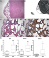
A–B. Renal xenograft (obtained at 20 weeks’ gestation, studied at post-transplant week 4) before (A) and after (B) laser capture microdissection of the region of plexus-forming angiogenesis. The remaining graft tissue is sampled in subsequent steps, and collected separately. The overlying renal (murine) capsule is not sampled. C–D. Higher magnification of fig. 4A illustrating the complex capillary network of plexus-forming angiogenesis with, in this case, apparent extension into the overlying renal capsule. A: Original magnification ×100; B: ×60, C–D: ×200. A, C: hematoxylin-eosin staining; D: CD-31 (PECAM-1) immunohistochemical analysis (DAB-peroxidase staining with hematoxylin counterstain). E. Results of qRT-PCR analysis of mRNA expression of IGF2, SULF2 and HMOX1 in graft regions without (“control”) or with (“PFA”) plexus-forming angiogenesis. Data represent mean ± SD. *: P < 0.05; **: P < 0.01 versus control.

Similar articles

Cited by

References

    1. Stoll BJ, Hansen NI, Bell EF, Shankaran S, Laptook AR, Walsh MC, Hale EC, Newman NS, Schibler K, Carlo WA, Kennedy KA, Poindexter BB, Finer NN, Ehrenkranz RA, Duara S, Sanchez PJ, O'Shea TM, Goldberg RN, Van Meurs KP, Faix RG, Phelps DL, Frantz ID, 3rd, Watterberg KL, Saha S, Das A, Higgins RD. Neonatal outcomes of extremely preterm infants from the NICHD Neonatal Research Network. Pediatrics. 2010;126:443–456. - PMC - PubMed
    1. Costeloe KL, Hennessy EM, Haider S, Stacey F, Marlow N, Draper ES. Short term outcomes after extreme preterm birth in England: comparison of two birth cohorts in 1995 and 2006 (the EPICure studies) BMJ. 2012;345:e7976. - PMC - PubMed
    1. Landry JS, Chan T, Lands L, Menzies D. Long-term impact of bronchopulmonary dysplasia on pulmonary function. Canadian respiratory journal : journal of the Canadian Thoracic Society. 2011;18:265–270. - PMC - PubMed
    1. Vom Hove M, Prenzel F, Uhlig HH, Robel-Tillig E. Pulmonary outcome in former preterm, very low birth weight children with bronchopulmonary dysplasia: a case-control follow-up at school age. J Pediatr. 2014;164:40–45. e44. - PubMed
    1. Husain AN, Siddiqui NH, Stocker JT. Pathology of arrested acinar development in postsurfactant bronchopulmonary dysplasia. Hum Pathol. 1998;29:710–717. - PubMed

Publication types

MeSH terms

LinkOut - more resources