

Heparin and LPS-induced COX-2 expression in airway cells: a link between its anti-inflammatory effects and GAG sulfation
- PMID: 26495958
- PMCID: PMC4962551
- DOI: 10.3109/01902148.2015.1091053
Heparin and LPS-induced COX-2 expression in airway cells: a link between its anti-inflammatory effects and GAG sulfation
Abstract
Purpose/aim: Previous studies have indicated that the sulfated polysaccharide heparin has anti-inflammatory effects. However, the mechanistic basis for these effects has not been fully elucidated.
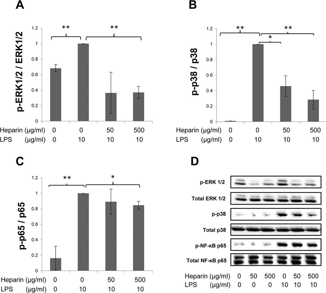
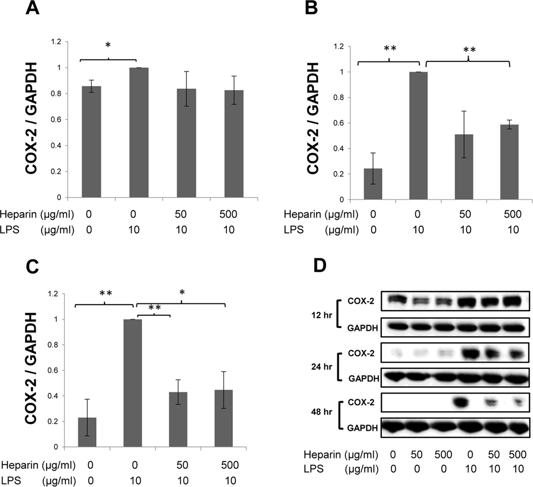
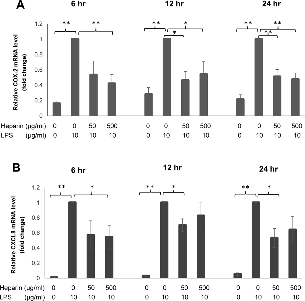
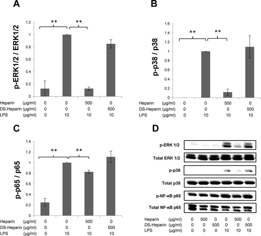
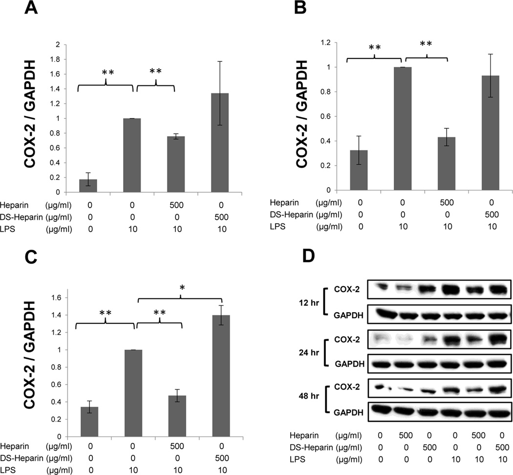
Materials and methods: NCI-H292 (mucoepidermoid) and HBE-1 (normal) human bronchial epithelial cells were treated with LPS alone or in the presence of high-molecular-weight (HMW) fully sulfated heparin or desulfated HMW heparin. Cells were harvested to examine the phosphorylation levels of ERK1/2, p38, and NF-kB p65 and COX-2 protein expression by Western blot and gene expression of both COX-2 and CXCL-8 by TaqMan qRT-PCR.
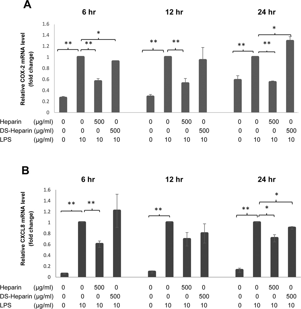
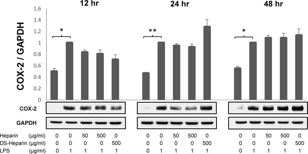
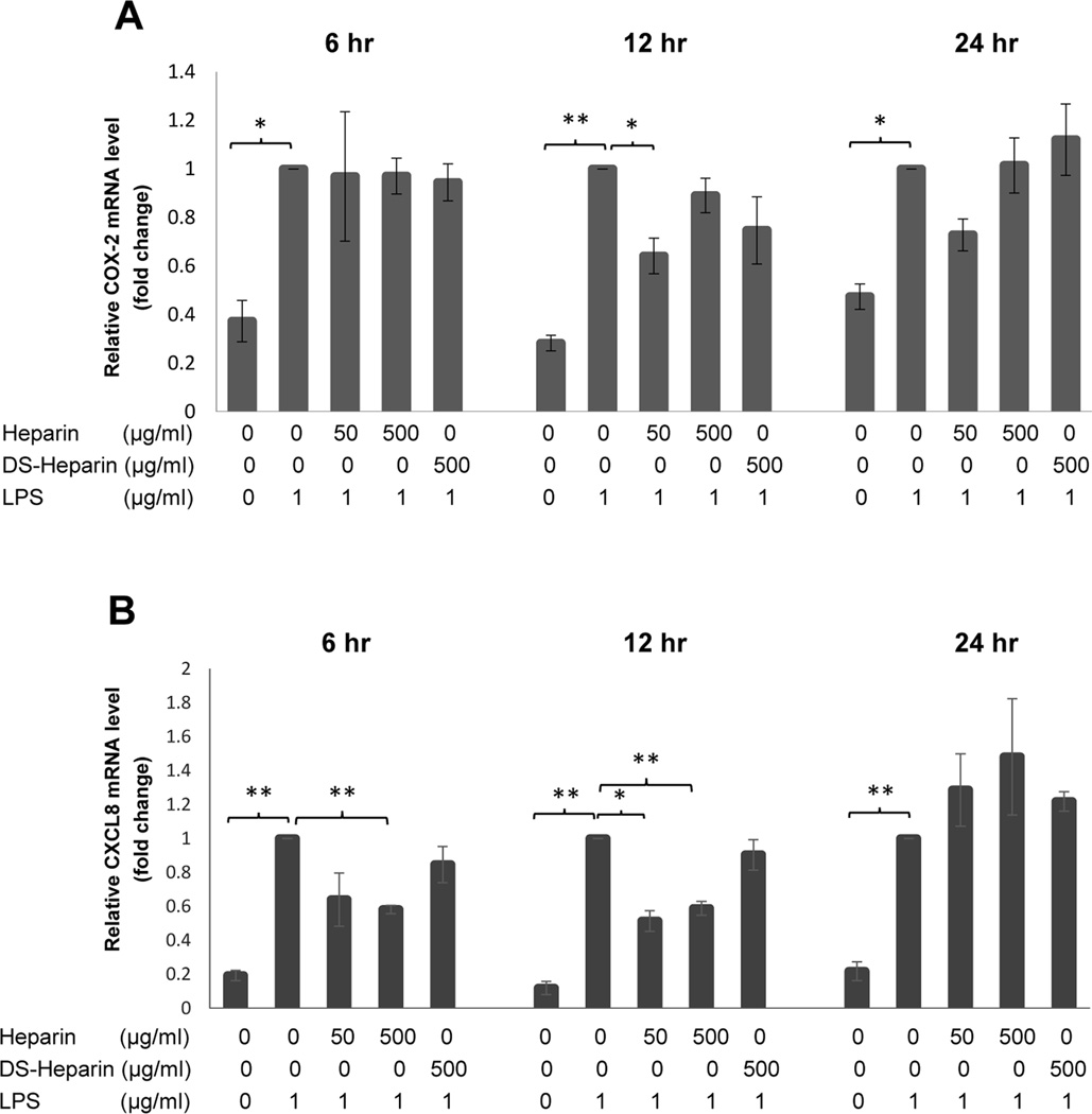
Results: Heparin is known to exert an influence on receptor-mediated signaling through its ability to both potentiate and inhibit the receptor-ligand interaction, depending upon its concentration. In H292 cells, fully-sulfated HMW heparin significantly reduced LPS-induced gene expression of both COX-2 and CXCL-8 for up to 48 hours, while desulfated heparin had little to no significant suppressive effect on signaling or on COX-2 gene or protein expression. Desulfated heparin, initially ineffective at preventing LPS-induced CXCL8 up-regulation, reduced CXCL8 transcription at 24 hours. In contrast, in normal HBE-1 cells, fully sulfated heparin significantly suppressed only ERK signaling, COX-2 gene expression at 12 hours, and CXCL-8 gene expression at 6 and 12 hours, while desulfated heparin had no significant effects on LPS-stimulated signaling or on gene or protein expression. Sulfation determines heparin's influence and may reflect the moderating role of GAG sulfation in lung injury and health.
Conclusions: Heparin's anti-inflammatory effects result from its nonspecific suppression of signaling and gene expression and are determined by its sulfation.
Keywords: COX-2; LPS; heparin; lung; sulfation.
Conflict of interest statement
No conflicts of interest, financial or otherwise
Figures

Similar articles
-
Phloretin inhibits interleukin-1β-induced COX-2 and ICAM-1 expression through inhibition of MAPK, Akt, and NF-κB signaling in human lung epithelial cells.Food Funct. 2015 Jun;6(6):1960-7. doi: 10.1039/c5fo00149h. Food Funct. 2015. PMID: 25996641
-
The liquid Panax ginseng inhibits epidermal growth factor-induced metalloproteinase 9 and cyclooxygenase 2 expressions via inhibition of inhibitor factor kappa-B-alpha and extracellular signal-regulated kinase in NCI-H292 human airway epithelial cells.Am J Rhinol Allergy. 2011 Mar-Apr;25(2):e55-9. doi: 10.2500/ajra.2011.25.3586. Am J Rhinol Allergy. 2011. PMID: 21679500
-
7b, a novel naphthalimide derivative, exhibited anti-inflammatory effects via targeted-inhibiting TAK1 following down-regulation of ERK1/2- and p38 MAPK-mediated activation of NF-κB in LPS-stimulated RAW264.7 macrophages.Int Immunopharmacol. 2013 Oct;17(2):216-28. doi: 10.1016/j.intimp.2013.06.008. Epub 2013 Jun 26. Int Immunopharmacol. 2013. PMID: 23810444
-
Structural requirements of heparin and related molecules to exert a multitude of anti-inflammatory activities.Mini Rev Med Chem. 2006 Sep;6(9):1009-23. doi: 10.2174/138955706778195180. Mini Rev Med Chem. 2006. PMID: 17018000 Review.
-
Engineering of routes to heparin and related polysaccharides.Appl Microbiol Biotechnol. 2012 Jan;93(1):1-16. doi: 10.1007/s00253-011-3641-4. Epub 2011 Nov 3. Appl Microbiol Biotechnol. 2012. PMID: 22048616 Free PMC article. Review.
Cited by
-
Role of Adaptor Protein Myeloid Differentiation 88 (MyD88) in Post-Subarachnoid Hemorrhage Inflammation: A Systematic Review.Int J Mol Sci. 2021 Apr 18;22(8):4185. doi: 10.3390/ijms22084185. Int J Mol Sci. 2021. PMID: 33919485 Free PMC article.
-
Repurposing High-Throughput Screening Reveals Unconventional Drugs with Antimicrobial and Antibiofilm Potential Against Methicillin-Resistant Staphylococcus aureus from a Cystic Fibrosis Patient.Antibiotics (Basel). 2025 Apr 14;14(4):402. doi: 10.3390/antibiotics14040402. Antibiotics (Basel). 2025. PMID: 40298549 Free PMC article.
-
Divergent Mast Cell Responses Modulate Antiviral Immunity During Influenza Virus Infection.Front Cell Infect Microbiol. 2021 Feb 19;11:580679. doi: 10.3389/fcimb.2021.580679. eCollection 2021. Front Cell Infect Microbiol. 2021. PMID: 33680987 Free PMC article. Review.
-
Pulmonary immunity and extracellular matrix interactions.Matrix Biol. 2018 Nov;73:122-134. doi: 10.1016/j.matbio.2018.04.003. Epub 2018 Apr 9. Matrix Biol. 2018. PMID: 29649546 Free PMC article. Review.
-
Mast Cells Exert Anti-Inflammatory Effects in an IL10-/- Model of Spontaneous Colitis.Mediators Inflamm. 2018 Apr 17;2018:7817360. doi: 10.1155/2018/7817360. eCollection 2018. Mediators Inflamm. 2018. PMID: 29849494 Free PMC article.
References
-
- Smits NC, Shworak NW, Dekhuijzen PN, van Kuppevelt TH. Heparan sulfates in the lung: structure, diversity, and role in pulmonary emphysema. Anat Rec. 2010;293(6):955–967. - PubMed
-
- Liang J, Jiang D, Griffith J, Yu S, Fan J, Zhao X, et al. CD44 is a negative regulator of acute pulmonary inflammation and lipopolysaccharide-TLR signaling in mouse macrophages. J Immunol. 2007;178(4):2469–2475. - PubMed
Publication types
MeSH terms
Substances
Grants and funding
LinkOut - more resources
Full Text Sources
Other Literature Sources
Medical
Research Materials
Miscellaneous