

A Metabolic Probe-Enabled Strategy Reveals Uptake and Protein Targets of Polyunsaturated Aldehydes in the Diatom Phaeodactylum tricornutum
- PMID: 26496085
- PMCID: PMC4619725
- DOI: 10.1371/journal.pone.0140927
A Metabolic Probe-Enabled Strategy Reveals Uptake and Protein Targets of Polyunsaturated Aldehydes in the Diatom Phaeodactylum tricornutum
Abstract
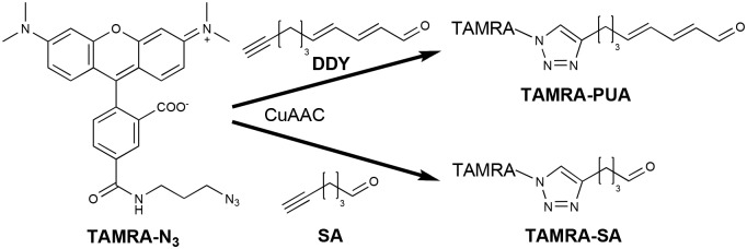
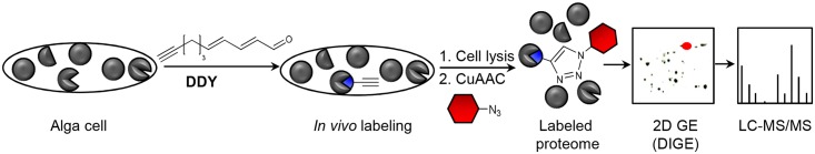
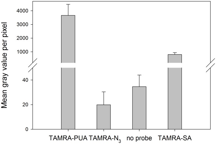
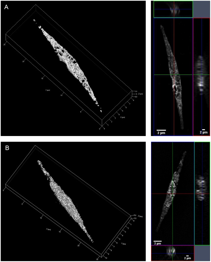
Diatoms are unicellular algae of crucial importance as they belong to the main primary producers in aquatic ecosystems. Several diatom species produce polyunsaturated aldehydes (PUAs) that have been made responsible for chemically mediated interactions in the plankton. PUA-effects include chemical defense by reducing the reproductive success of grazing copepods, allelochemical activity by interfering with the growth of competing phytoplankton and cell to cell signaling. We applied a PUA-derived molecular probe, based on the biologically highly active 2,4-decadienal, with the aim to reveal protein targets of PUAs and affected metabolic pathways. By using fluorescence microscopy, we observed a substantial uptake of the PUA probe into cells of the diatom Phaeodactylum tricornutum in comparison to the uptake of a structurally closely related control probe based on a saturated aldehyde. The specific uptake motivated a chemoproteomic approach to generate a qualitative inventory of proteins covalently targeted by the α,β,γ,δ-unsaturated aldehyde structure element. Activity-based protein profiling revealed selective covalent modification of target proteins by the PUA probe. Analysis of the labeled proteins gave insights into putative affected molecular functions and biological processes such as photosynthesis including ATP generation and catalytic activity in the Calvin cycle or the pentose phosphate pathway. The mechanism of action of PUAs involves covalent reactions with proteins that may result in protein dysfunction and interference of involved pathways.
Conflict of interest statement
Figures
Similar articles
-
Accumulation of polyunsaturated aldehydes in the gonads of the copepod Acartia tonsa revealed by tailored fluorescent probes.PLoS One. 2014 Nov 10;9(11):e112522. doi: 10.1371/journal.pone.0112522. eCollection 2014. PLoS One. 2014. PMID: 25383890 Free PMC article.
-
Quantification of dissolved and particulate polyunsaturated aldehydes in the Adriatic sea.Mar Drugs. 2011;9(4):500-513. doi: 10.3390/md9040500. Epub 2011 Mar 29. Mar Drugs. 2011. PMID: 21731545 Free PMC article.
-
Survey of the chemical defence potential of diatoms: screening of fifty one species for alpha,beta,gamma,delta-unsaturated aldehydes.J Chem Ecol. 2005 Apr;31(4):949-58. doi: 10.1007/s10886-005-3615-z. J Chem Ecol. 2005. PMID: 16124261
-
Diatom/copepod interactions in plankton: the indirect chemical defense of unicellular algae.Chembiochem. 2005 Jun;6(6):946-59. doi: 10.1002/cbic.200400348. Chembiochem. 2005. PMID: 15883976 Review.
-
Toxigenic effects of diatoms on grazers, phytoplankton and other microbes: a review.Ecotoxicology. 2010 Mar;19(3):493-511. doi: 10.1007/s10646-009-0434-y. Ecotoxicology. 2010. PMID: 19924531 Review.
References
-
- Nelson DM, Tréguer P, Brzezinski MA, Leynaert A, Quéguiner B. Production and dissolution of biogenic silica in the ocean: Revised global estimates, comparison with regional data and relationship to biogenic sedimentation. Global Biogeochem Cycles. 1995;9(3):359–72.
-
- Pohnert G. Diatom/Copepod interactions in plankton: The Indirect chemical defense of unicellular algae. ChemBioChem. 2005;6(6):946–59. - PubMed
Publication types
MeSH terms
Substances
LinkOut - more resources
Full Text Sources
Other Literature Sources
