

Modular Coils with Low Hydrogen Content Especially for MRI of Dry Solids
- PMID: 26496192
- PMCID: PMC4619699
- DOI: 10.1371/journal.pone.0139763
Modular Coils with Low Hydrogen Content Especially for MRI of Dry Solids
Erratum in
-
Correction: Modular Coils with Low Hydrogen Content Especially for MRI of Dry Solids.PLoS One. 2016 Mar 9;11(3):e0151505. doi: 10.1371/journal.pone.0151505. eCollection 2016. PLoS One. 2016. PMID: 26960147 Free PMC article. No abstract available.
Abstract
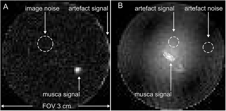
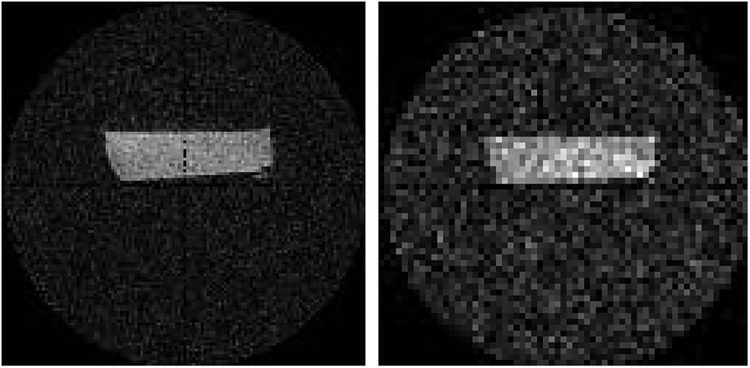
Introduction: Recent advances have enabled fast magnetic resonance imaging (MRI) of solid materials. This development has opened up new applications for MRI, but, at the same time, uncovered new challenges. Previously, MRI-invisible materials like the housing of MRI detection coils are now readily depicted and either cause artifacts or lead to a decreased image resolution. In this contribution, we present versatile, multi-nuclear single and dual-tune MRI coils that stand out by (1) a low hydrogen content for high-resolution MRI of dry solids without artifacts; (2) a modular approach with exchangeable inductors of variable volumes to optimally enclose the given object; (3) low cost and low manufacturing effort that is associated with the modular approach; (4) accurate sample placement in the coil outside of the bore, and (5) a wide, single- or dual-tune frequency range that covers several nuclei and enables multinuclear MRI without moving the sample.
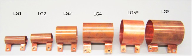
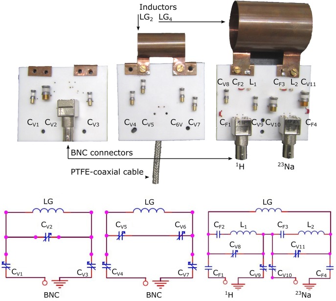
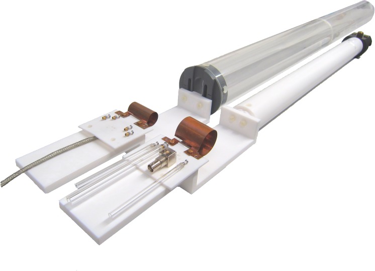
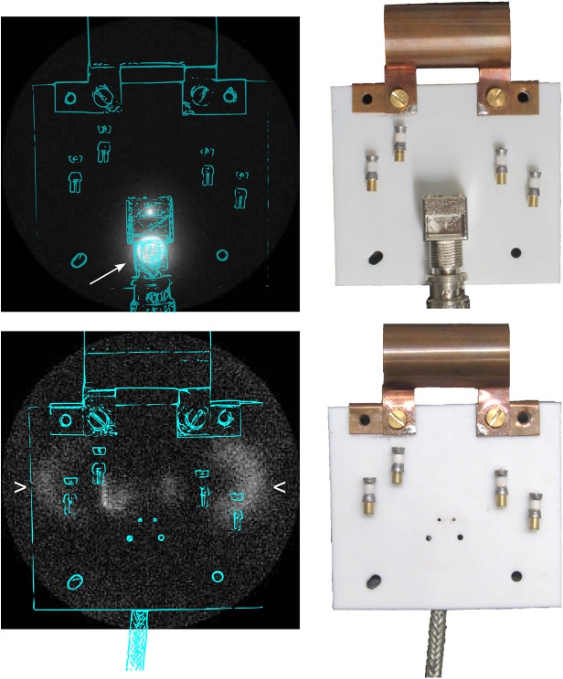
Materials and methods: The inductors of the coils were constructed from self-supporting copper sheets to avoid all plastic materials within or around the resonator. The components that were mounted at a distance from the inductor, including the circuit board, coaxial cable and holder were manufactured from polytetrafluoroethylene.
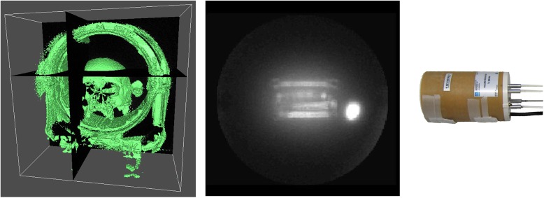
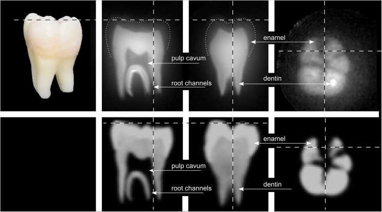
Results and conclusion: Residual hydrogen signal was sufficiently well suppressed to allow 1H-MRI of dry solids with a minimum field of view that was smaller than the sensitive volume of the coil. The SNR was found to be comparable but somewhat lower with respect to commercial, proton-rich quadrature coils, and higher with respect to a linearly-polarized commercial coil. The potential of the setup presented was exemplified by 1H/23Na high-resolution zero echo time (ZTE) MRI of a model solution and a dried human molar at 9.4 T. A full 3D image dataset of the tooth was obtained, rich in contrast and similar to the resolution of standard cone-beam computed tomography.
Conflict of interest statement
Figures
Similar articles
-
A PIN diode controlled dual-tuned MRI RF coil and phased array for multi nuclear imaging.Phys Med Biol. 2010 May 7;55(9):2589-600. doi: 10.1088/0031-9155/55/9/011. Epub 2010 Apr 14. Phys Med Biol. 2010. PMID: 20393229
-
A virtually 1H-free birdcage coil for zero echo time MRI without background signal.Magn Reson Med. 2017 Jul;78(1):399-407. doi: 10.1002/mrm.26368. Epub 2016 Aug 9. Magn Reson Med. 2017. PMID: 27505183
-
Rapid and robust pulmonary proton ZTE imaging in the mouse.NMR Biomed. 2014 Sep;27(9):1129-34. doi: 10.1002/nbm.3161. Epub 2014 Jul 26. NMR Biomed. 2014. PMID: 25066371
-
A decoupled coil detector array for fast image acquisition in magnetic resonance imaging.Med Phys. 1991 Mar-Apr;18(2):251-65. doi: 10.1118/1.596723. Med Phys. 1991. PMID: 2046612
-
Intracranial microvascular imaging at 7 T MRI with transceiver RF coils.Magn Reson Imaging. 2014 Nov;32(9):1133-8. doi: 10.1016/j.mri.2014.07.006. Epub 2014 Aug 2. Magn Reson Imaging. 2014. PMID: 25093629
Cited by
-
Imaging of root canal treatment using ultra high field 9.4T UTE-MRI - a preliminary study.Dentomaxillofac Radiol. 2020 Jan;49(1):20190183. doi: 10.1259/dmfr.20190183. Epub 2019 Sep 23. Dentomaxillofac Radiol. 2020. PMID: 31530016 Free PMC article.
-
Dental MRI using wireless intraoral coils.Sci Rep. 2016 Mar 29;6:23301. doi: 10.1038/srep23301. Sci Rep. 2016. PMID: 27021387 Free PMC article.
-
Correction: Modular Coils with Low Hydrogen Content Especially for MRI of Dry Solids.PLoS One. 2016 Mar 9;11(3):e0151505. doi: 10.1371/journal.pone.0151505. eCollection 2016. PLoS One. 2016. PMID: 26960147 Free PMC article. No abstract available.
References
-
- Weiger M, Pruessmann KP. MRI with Zero Echo Time. eMagRes. 2012;1: 311–322. 10.1002/9780470034590.emrstm1292 - DOI
-
- Garwood M, Idiyatullin D, Corum CA, Chamberlain R, Moeller S, Kobayashi N, et al. Capturing Signals from Fast-relaxing Spins with Frequency-Swept MRI: SWIFT. eMagRes. 2012;1 10.1002/9780470034590.emrstm1259 - DOI
-
- Emid S, Creyghton JHN. High resolution NMR imaging in solids. Physica B+C. 1985;128: 81–83. 10.1016/0378-4363(85)90087-7 - DOI
-
- Gravina S, Cory DG. Sensitivity and Resolution of Constant-Time Imaging. Journal of Magnetic Resonance, Series B. 1994;104: 53–61. 10.1006/jmrb.1994.1052 - DOI
Publication types
MeSH terms
Substances
LinkOut - more resources
Full Text Sources
Other Literature Sources
Medical
