Observational Study
. 2015 Oct;94(42):e1667.
doi: 10.1097/MD.0000000000001667.

Emergency Transcatheter Arterial Embolization for Acute Renal Hemorrhage

Affiliations
Observational Study

Emergency Transcatheter Arterial Embolization for Acute Renal Hemorrhage

Hong Liang Wang et al. Medicine (Baltimore). 2015 Oct.

Abstract

The aims of this study were to identify arteriographic manifestations of acute renal hemorrhage and to evaluate the efficacy of emergency embolization. Emergency renal artery angiography was performed on 83 patients with acute renal hemorrhage. As soon as bleeding arteries were identified, emergency embolization was performed using gelatin sponge, polyvinyl alcohol particles, and coils. The arteriographic presentation and the effect of the treatment for acute renal hemorrhage were analyzed retrospectively. Contrast extravasation was observed in 41 patients. Renal arteriovenous fistulas were found in 12 of the 41 patients. In all, 8 other patients had a renal pseudoaneurysm, 5 had pseudoaneurysm rupture complicated by a renal arteriovenous fistula, and 1 had pseudoaneurysm rupture complicated by a renal artery-calyceal fistula. Another 16 patients had tumor vasculature seen on arteriography. Before the procedure, 35 patients underwent renal artery computed tomography angiography (CTA). Following emergency embolization, complete hemostasis was achieved in 80 patients, although persistent hematuria was present in 3 renal trauma patients and 1 patient who had undergone percutaneous nephrolithotomy (justifying surgical removal of the ipsilateral kidney in this patient). Two-year follow-up revealed an overall effective rate of 95.18 % (79/83) for emergency embolization. There were no serious complications. Emergency embolization is a safe, effective, minimally invasive treatment for renal hemorrhage. Because of the diversified arteriographic presentation of acute renal hemorrhage, proper selection of the embolic agent is a key to successful hemostasis. Preoperative renal CTA plays an important role in diagnosing and localizing the bleeding artery.


Conflict of interest statement

There are no conflicts of interest in this study.

Figures
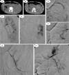
FIGURE 1
FIGURE 1
A 14-year-old boy who had undergone surgical resection of the right kidney presented with persistent hematuria after surgery for left renal trauma. (A, B) Renal CT images before embolization show that the left kidney is enlarged, the capsule is not intact, and a high-density hematoma is obvious in the renal pelvis and hilum. (C, D) Angiography with a Cobra catheter and microcatheter reveal contrast extravasation in the lower branch of the left renal artery. (E) Microcatheter angiography following polyvinyl alcohol (PVA) (300–500 μm) embolization. The previously bleeding artery is successfully occluded. (F) Contrast extravasation of the posterior branch of the left renal artery. (G) Cobra angiography following PVA embolization (300–500 μm). The previously bleeding artery has been successfully occluded. (H) Angiography with the Cobra catheter. All bleeding arteries have been completely occluded. Normal renal arteries and the renal parenchyma are visible.
FIGURE 2
FIGURE 2
A 38-year-old man presented with persistent massive hematuria after trauma to the right kidney. (A) Angiography with the PIG catheter showed bilateral renal arteries. The left kidney appears to be normal, whereas the trunk of the right renal artery is thin, and the right renal parenchyma is unclear. (B) Microcatheter angiography reveals a pseudoaneurysm of the upper branch of the right renal artery (black arrow). (C) Polyvinyl alcohol particles (300–500 μm) angiography after embolization. The pseudoaneurysm has disappeared, and normal renal artery and renal parenchyma are visible.
FIGURE 3
FIGURE 3
A 38-year-old man had persistent massive hematuria due to a closed contusion of the right kidney in a car accident. (A) Renal artery computed tomography angiography shows a pseudoaneurysm in the pelvis of the lower pole of the right kidney (white arrow). (B) Enhanced CT shows subcapsular effusion of the right kidney with an aneurysm-like enlargement of the right pelvis (white arrow). (C) Angiography with the Cobra catheter shows that the pseudoaneurysm in the lower pole of the right kidney is ruptured and bleeding (long black arrow), and there is contrast extravasation (short black arrow). An arteriovenous fistula has formed in the right kidney, and early opacification of the inferior vena cava is seen (white arrow). (D) Angiography with the Cobra catheter (after embolization with 2 MWCE35-3-3 coils and 1 gelatin sponge strip) shows complete occlusion of the bleeding artery. The pseudoaneurysm and renal arteriovenous fistula are not visible. The surrounding kidney tissues appear normal.
FIGURE 4
FIGURE 4
A 35-year-old woman presented with sustained hematuria after a right kidney biopsy. (A) Cobra angiography shows a pseudoaneurysm of the small branch of the posterior segment of the right renal artery (black arrow). (B) Superselective angiography reveals early opacification of the right renal pelvis and the ureter (black arrow) at the arterial phase (renal artery–renal pelvis fistula). (C) Repeat polyvinyl alcohol particles (500–710 μm) angiography after embolization shows that the bleeding artery is completely occluded with no sign of contrast extravasation. The surrounding kidney tissues appear normal.
FIGURE 5
FIGURE 5
A 58-year-old man with a right renal tumor presented with persistent massive hematuria. (A) Renal artery computed tomography angiography reveals that the right renal artery (long white arrow) and the right accessory renal artery (short white arrow) are feeding the tumor. (B, C) Angiography with the Cobra catheter and microcatheter show an artery feeding the tumor in the upper pole of the right kidney. A superselective microcatheter is placed in the feeding artery for embolization. (D) Polyvinyl alcohol (PVA) particles (300–500 μm) angiography after embolization shows that the artery feeding the tumor in the upper pole of the right kidney is successfully occluded. Normal kidney tissues of the lower pole are visible. (E) Superselective microcatheter placement in the accessory renal artery (white arrow) shows the right tumor artery. (F) Microcatheter angiography following PVA particles (300–500 μm) embolization shows that the feeding artery is completely occluded. Hematuria disappeared after the surgery, and the removed tumor was pathologically confirmed as cell carcinoma of the right kidney.

References

    1. Sommer CM, Stampfl U, Bellemann N, et al. Patients with life-threatening arterial renal hemorrhage: CT angiography and catheter angiography with subsequent superselective embolization. Cardiovasc Intervent Radiol 2010; 33:498–508. - PubMed
    1. Zugazagoitia J, Sastre J, Barrera J, et al. Severe perirenal hematoma in a patient with a single kidney treated with sunitinib for metastatic pancreatic neuroendocrine tumor. J Cancer Res Ther 2012; 8:303–305. - PubMed
    1. Springer C, Hoda MR, Fajkovic H, et al. Laparoscopic vs open partial nephrectomy for T1 renal tumours: evaluation of long-term oncological and functional outcomes in 340 patients. BJU Int 2013; 111:281–288. - PubMed
    1. Keeling AN, McGrath FP, Thornton J, et al. Emergency percutaneous transcatheter embolisation of acute arterial haemorrhage. Ir J Med Sci 2010; 179:385–391. - PubMed
    1. Koganemaru M, Abe T, Iwamoto R, et al. Ultraselective arterial embolization of vasa recta using 1.7-French microcatheter with small-sized detachable coils in acute colonic hemorrhage after failed endoscopic treatment. Am J Roentgenol 2012; 198:W370–372. - PubMed

Publication types