

Branchial Cleft-Like Cysts Involving 3 Different Organs: Thyroid Gland, Thymus, and Parotid Gland
- PMID: 26496296
- PMCID: PMC4620827
- DOI: 10.1097/MD.0000000000001758
Branchial Cleft-Like Cysts Involving 3 Different Organs: Thyroid Gland, Thymus, and Parotid Gland
Abstract
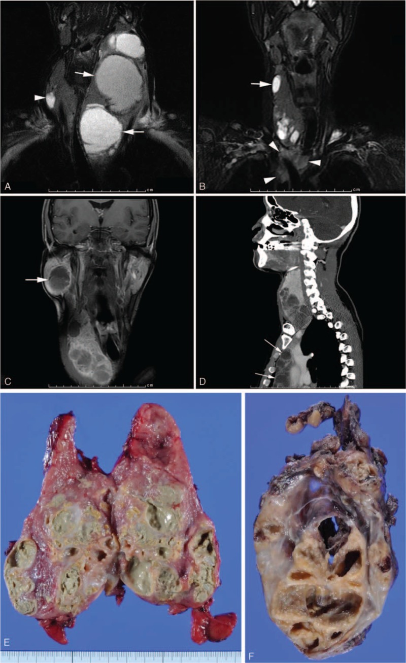
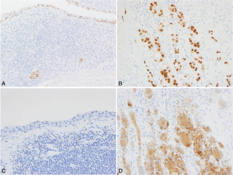
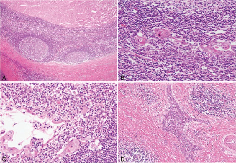
Branchial cleft cysts (BCCs) are also named lateral cervical cysts and widely acknowledged as being derived from embryonic remnants. Lymphoepithelial cysts (LECs) generally show microscopic features that are identical to those of BCCs, and rarely occur at unusual sites or organs.A case of multiple cysts arising in both lobes of the thyroid gland, thymus, and right parotid gland in a 41-year-old man is reported. Clinically, the patient presented with Hashimoto's thyroiditis for about 20 years and had past histories of idiopathic thrombocytopenic purpura and severe respiratory infection.This case is unusual in that multiple cysts arose synchronously and/or heterochronously and grew, increasing their sizes in these different organs. Microscopic examinations revealed that all of the cysts were composed of squamous epithelium, dense lymphoid tissue with germinal centers, and a fibrous capsule. These findings corresponded to those of BCCs or LECs. It is notable that the histopathological features were nearly the same in the individual organs. A review of the literature disclosed no previous such reported cases.The etiology is unknown. However, based upon the similar histopathological features of all the excised specimens, common immune and/or hematopoietic disorders may have contributed to their occurrence and development in association with putative genetic abnormalities.
Conflict of interest statement
The authors have no funding and conflicts of interest to disclose.
Figures


Similar articles
-
Intrathyroidal lymphoepithelial cysts of probable branchial origin.Hum Pathol. 1994 Nov;25(11):1238-42. doi: 10.1016/0046-8177(94)90042-6. Hum Pathol. 1994. PMID: 7959670
-
Multiple branchial cleft-like cysts in Hashimoto's thyroiditis.Am J Surg Pathol. 1989 Jan;13(1):45-9. doi: 10.1097/00000478-198901000-00006. Am J Surg Pathol. 1989. PMID: 2909197
-
Synchronous Presentation of Right Parotid Branchial Cleft Cyst and Left Neck HPV-Associated Cystic Metastatic Squamous Cell Carcinoma: A Diagnostic Quandary.Ear Nose Throat J. 2025 Mar;104(1_suppl):420S-423S. doi: 10.1177/01455613231158803. Epub 2023 Feb 16. Ear Nose Throat J. 2025. PMID: 36798986
-
Primary papillary carcinoma of the thyroid arising in a branchial cyst: case report and review of the literature.Ear Nose Throat J. 2013 Feb;92(2):E3-5. Ear Nose Throat J. 2013. PMID: 23460225 Review.
-
Lymphoepithelial cysts of the thyroid gland. A case report and review of the literature.Arch Pathol Lab Med. 2003 Apr;127(4):e205-8. doi: 10.5858/2003-127-e205-LCOTTG. Arch Pathol Lab Med. 2003. PMID: 12683903 Review.
Cited by
-
Internal Hypopharyngeal Cyst: A Review of Literature.Dysphagia. 2019 Aug;34(4):487-498. doi: 10.1007/s00455-019-10003-2. Epub 2019 Mar 29. Dysphagia. 2019. PMID: 30927081 Review.
-
Cystic lesions of the parotid gland: radiologic-pathologic correlation according to the latest World Health Organization 2017 Classification of Head and Neck Tumours.Jpn J Radiol. 2017 Nov;35(11):629-647. doi: 10.1007/s11604-017-0678-z. Epub 2017 Aug 23. Jpn J Radiol. 2017. PMID: 28836142 Review.
References
-
- Coleman WR, Homer RS, Kaplan RP. Branchial cleft heterotopia of the lower neck. J Cutan Pathol 1989; 16:353–358. - PubMed
-
- Giuta J, Cantaldo E. Lymphoepithelial cysts of the oral mucosa. Oral Surg Oral Med Oral Pathol 1973; 35:77–84. - PubMed
-
- Ihrler S, Zietz C, Riederer A, et al. HIV-related parotid lymphoepithelial cysts. Immunohistochemistry and 3-D reconstruction of surgical and autopsy material with special reference to formal pathogenesis. Virchows Arch 1996; 429:13–47. - PubMed
-
- Truong LD, Rangdaeng S, Jordan PH. Lymphoepithelial cyst of the pancreas. Am J Surg Pathol 1987; 11:899–903. - PubMed
Publication types
MeSH terms
LinkOut - more resources
Full Text Sources
Medical
Miscellaneous
