Arthroscopic Resection of a Tenosynovial Giant Cell Tumor in the Wrist: A Case Report
- PMID: 26496348
- PMCID: PMC4620757
- DOI: 10.1097/MD.0000000000001887
Arthroscopic Resection of a Tenosynovial Giant Cell Tumor in the Wrist: A Case Report
Abstract
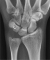
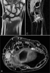
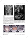
The treatment for giant cell tumors of the tendon sheath is surgical therapy, but surgical recurrence rates were reported to be as high as 50% in some cases. Therefore, complete radical excision of the lesion is the treatment of choice. If the tumor originates from the joint, it is important to perform capsulotomy. Here, the authors report the first case of successful treatment of a localized intra-articular giant cell tumor in the wrist by arthroscopic resection.A 28-year-old right-handed woman visited the clinic because of left wrist ulnar-side pain, which had been aggravated during the previous 15 days. Vague ulnar-side wrist pain had begun 2 years ago. When the authors examined the patient, the wrist showed mild swelling on the volo-ulnar aspect and the distal radioulnar joint, as well as volar joint line tenderness. She showed a positive result on the ulnocarpal stress test and displayed limited range of motion. Magnetic resonance imaging revealed an intra-articular mass with synovitis in the ulnocarpal joint. Wrist arthroscopy was performed using standard portals under regional anesthesia. The arthroscopic findings revealed a large, well-encapsulated, yellow lobulated soft-tissue mass that was attached to the volar side of the ulnocarpal ligament and connected to the extra-articular side. The mass was completely excised piece by piece with a grasping forceps. Histopathologic examination revealed that the lesion was an intra-articular localized form of a tenosynovial giant cell tumor.At 24-month follow-up, the patient was completely asymptomatic and had full range of motion in her left wrist, and no recurrence was found in magnetic resonance imaging follow-up evaluations.The authors suggest that the arthroscopic excision of intra-articular giant cell tumors, as in this case, may be an alternative method to open excisions, with many advantages.
Conflict of interest statement
The authors have no conflicts of interest to disclose.
Figures


Similar articles
-
Intra-articular tenosynovial giant cell tumor arising from the posterior cruciate ligament.Orthopedics. 2012 Jul 1;35(7):e1116-8. doi: 10.3928/01477447-20120621-34. Orthopedics. 2012. PMID: 22784912
-
Arthroscopic resection of an extra-articular tenosynovial giant cell tumor from the ankle region.Arthroscopy. 2003 Sep;19(7):E8-11. doi: 10.1016/s0749-8063(03)00687-x. Arthroscopy. 2003. PMID: 12966401
-
Distinct extra-articular invasion patterns of diffuse pigmented villonodular synovitis/tenosynovial giant cell tumor in the knee joints.Knee Surg Sports Traumatol Arthrosc. 2018 Nov;26(11):3508-3514. doi: 10.1007/s00167-018-4942-2. Epub 2018 Apr 10. Knee Surg Sports Traumatol Arthrosc. 2018. PMID: 29637236
-
Extra-articular localized nodular synovitis (giant cell tumor of tendon sheath origin) attached to the subtalar joint.Foot Ankle Int. 1996 Jul;17(7):413-6. doi: 10.1177/107110079601700710. Foot Ankle Int. 1996. PMID: 8832249 Review.
-
Giant cell tumor of tendon sheath, tenosynovial giant cell tumor, and pigmented villonodular synovitis: defining the presentation, surgical therapy and recurrence.Oncol Rep. 2000 Mar-Apr;7(2):413-9. Oncol Rep. 2000. PMID: 10671695 Review.
Cited by
-
Don't Dismiss the Swelling: A Rare Case of Tenosynovial Giant Cell Tumor in a Young Woman's Finger - A Case Report.J Orthop Case Rep. 2025 Aug;15(8):231-234. doi: 10.13107/jocr.2025.v15.i08.5950. J Orthop Case Rep. 2025. PMID: 40786784 Free PMC article.
-
Pigmented Villonodular Synovitis as an Atypical Cause of Deep Motor Branch Neuropathy.J Orthop Case Rep. 2021 Apr;11(4):80-84. doi: 10.13107/jocr.2021.v11.i04.2162. J Orthop Case Rep. 2021. PMID: 34327172 Free PMC article.
-
A New Simple and Practical Clinical Classification for Tenosynovial Giant Cell Tumors of the Knee.Orthop Surg. 2022 Feb;14(2):290-297. doi: 10.1111/os.13179. Epub 2021 Dec 16. Orthop Surg. 2022. PMID: 34914180 Free PMC article.
References
-
- Kotwal PP, Gupta V, Malhotra R. Giant-cell tumor of the tendon sheath. Is radiotherapy indicated to prevent recurrence after surgery? J Bone Joint Surg Br 2000; 82:571–573. - PubMed
-
- Moore JR, Weiland AJ, Curtis RM. Localized nodular tenosynovitis: experience with 115 cases. J Hand Surg Am 1984; 9:412–417. - PubMed
-
- Glowacki KA, Weiss APC. Giant cell tumors of tendon sheath. Hand Clin 1995; 11:245–253. - PubMed
-
- Nuahra ME, Bucchieri JS. Ganglion cysts and other tumor related conditions of the hand and wrist. Hand Clin 2004; 20:249–260. - PubMed
Publication types
MeSH terms
LinkOut - more resources
Full Text Sources

