. 2015 Dec 15;87(24):12032-9.
doi: 10.1021/acs.analchem.5b02087. Epub 2015 Nov 19.

Timescales and Frequencies of Reversible and Irreversible Adhesion Events of Single Bacterial Cells

Affiliations

Timescales and Frequencies of Reversible and Irreversible Adhesion Events of Single Bacterial Cells

Michelle D Hoffman et al. Anal Chem. .

Abstract

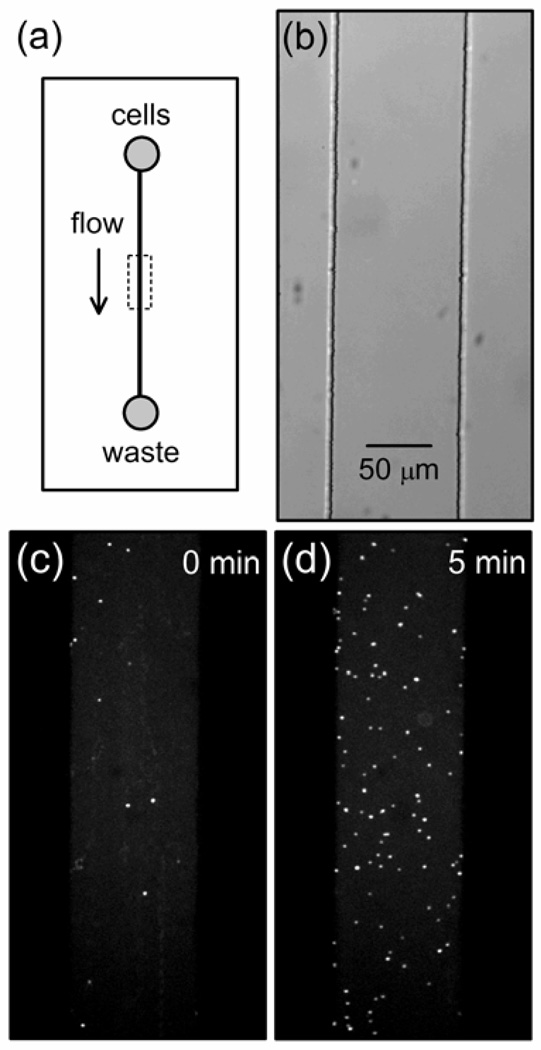
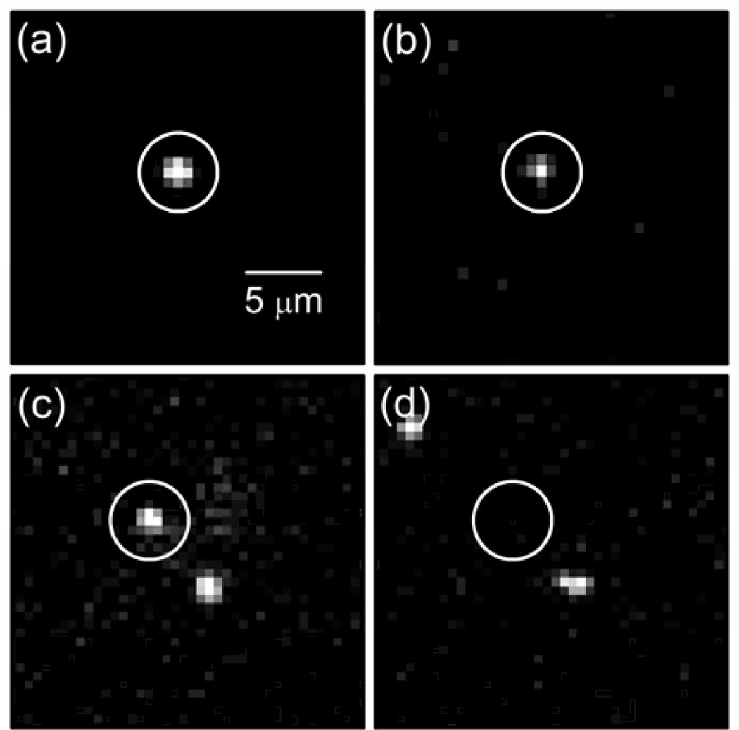
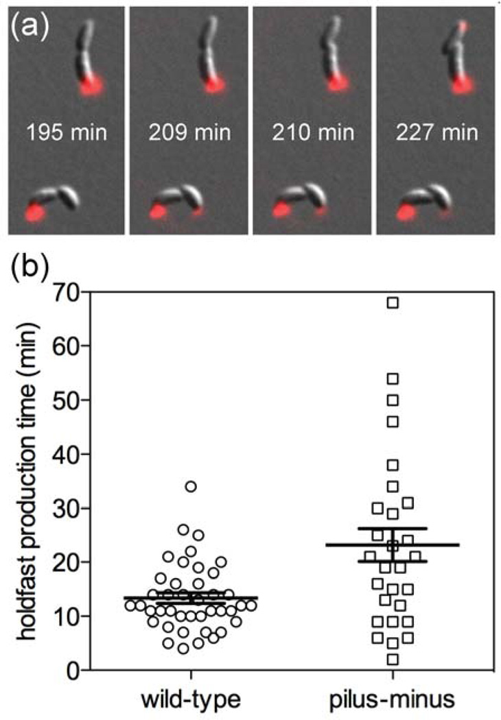
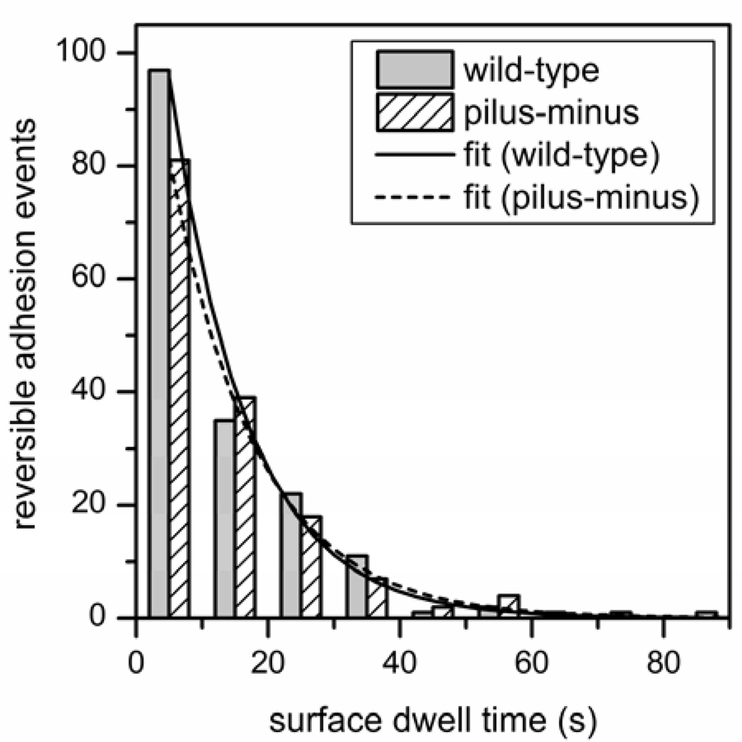
In the environment, most bacteria form surface-attached cell communities called biofilms. The attachment of single cells to surfaces involves an initial reversible stage typically mediated by surface structures such as flagella and pili, followed by a permanent adhesion stage usually mediated by polysaccharide adhesives. Here, we determine the absolute and relative timescales and frequencies of reversible and irreversible adhesion of single cells of the bacterium Caulobacter crescentus to a glass surface in a microfluidic device. We used fluorescence microscopy of C. crescentus expressing green fluorescent protein to track the swimming behavior of individual cells prior to adhesion, monitor the cell at the surface, and determine whether the cell reversibly or irreversibly adhered to the surface. A fluorescently labeled lectin that binds specifically to polar polysaccharides, termed holdfast, discriminated irreversible adhesion events from reversible adhesion events where no holdfast formed. In wild-type cells, the holdfast production time for irreversible adhesion events initiated by surface contact (23 s) was 30-times faster than the holdfast production time that occurs through developmental regulation (13 min). Irreversible adhesion events in wild-type cells (3.3 events/min) are 15-times more frequent than in pilus-minus mutant cells (0.2 events/min), indicating the pili are critical structures in the transition from reversible to irreversible surface-stimulated adhesion. In reversible adhesion events, the dwell time of cells at the surface before departing was the same for wild-type cells (12 s) and pilus-minus mutant cells (13 s), suggesting the pili do not play a significant role in reversible adhesion. Moreover, reversible adhesion events in wild-type cells (6.8 events/min) occur twice as frequently as irreversible adhesion events (3.3 events/min), demonstrating that most cells contact the surface multiple times before transitioning from reversible to irreversible adhesion.


Figures

Figure 1
Figure 1
(a) Schematic of a microfluidic device with a single poly(dimethylsiloxane) (PDMS) microchannel sealed with a glass cover plate. To deliver bacteria to the analysis region (dashed box), hydrostatic flow is generated by a higher fluid level in the cell reservoir relative to the waste reservoir. (b) Bright-field image of a 100-µm wide microchannel cast in PDMS with a glass cover plate. (c)–(d) Fluorescence images of the GFP signal from wild-type C. crescentus cells adhered to the glass cover plate at (c) 0 and (d) 5 min after the start of the experiment. The images were averaged for 5 s (or 20 frames) to minimize signal from the motile swarmer cells.
Figure 2
Figure 2
(a) GFP fluorescence image of an irreversible (permanent) adhesion event of a wild-type cell at the glass surface and (b) fluorescence image of labeled lectin (WGA-AF555) bound to the holdfast of the cell in panel (a). The holdfast production time for the cell was 19 s. GFP fluorescence images of (c) a reversible adhesion event of wild-type cell at the glass surface and (d) the same location as panel (c) after the cell detached from the surface. The dwell time for the cell was 12 s. Images in panels (a)–(d) were acquired at 20× magnification and have the same scale, and the white circles highlight the events described.
Figure 3
Figure 3
Histogram of holdfast production time after wild-type cells adhere to the glass surface. The holdfast production time is the time needed for the cell to secrete (or synthesize) holdfast after initially adhering to the surface. The histogram includes only cells that were motile prior to irreversible adhesion. Total number of irreversible adhesion events is 631, and the bin size is 10 s. The fitted line is a single exponential decay with a time constant for the holdfast production time of 23.1 ± 1.1 s.
Figure 4
Figure 4
Holdfast production time on agarose pads. (a) Composite images from DIC and fluorescence microscopy of wild-type cells used to determine the time of cell division (195 min for the cell on the bottom; 210 min for the cell on the top) and the time of holdfast detection (209 min for the cell on the bottom; 227 min for the cell on the top). The holdfast production time is the time of holdfast detection minus the time of cell division. (b) Holdfast production times for wild-type cells and pilus-minus mutant cells on agarose pads. The average times and standard deviations for holdfast production, indicated by the bars, are 13.4 ± 1.0 min for 41 wild-type cells and 23.2 ± 3.1 min for 28 pilus-minus mutant cells. For these experiments, agarose pads, instead of microfluidic devices, were used to minimize polar contact and surface contact stimulation.
Figure 5
Figure 5
Histograms of dwell times at the surface for wild-type cells and the pilus-minus mutant cells. Dwell time is the time of a reversible adhesion event during which a cell adheres to the surface, resides without forming a holdfast, and then detaches. Total numbers of reversible adhesion events are 171 for the wild-type cells and 159 for the pilus-minus mutants, and the bin size is 10 s. Fitted lines are single exponential decays with time constants of 11.7 ± 0.5 s for the wild-type cells and 13.2 ± 0.3 s for the pilus-minus mutant cells.

References

    1. Karatan E, Watnick P. Microbiol. Mol. Biol. Rev. 2009;73:310–347. - PMC - PubMed
    1. Potera C. Science. 1999;283:1837–1839. - PubMed
    1. Sintim HO, Smith JAI, Wang J, Nakayama S, Yan L. Future Med. Chem. 2010;2:1005–1035. - PubMed
    1. Cegelski L, Marshall GR, Eldridge GR, Hultgren SJ. Nat. Rev. Microbiol. 2008;6:17–27. - PMC - PubMed
    1. Rasko DA, Sperandio V. Nat. Rev. Drug Discov. 2010;9:117–128. - PubMed

Publication types

Substances

LinkOut - more resources