Multicenter Study
. 2015 Nov;205(5):1068-74.
doi: 10.2214/AJR.14.13330.

Hippocampal Malrotation Is Associated With Prolonged Febrile Seizures: Results of the FEBSTAT Study

Collaborators, Affiliations
Multicenter Study

Hippocampal Malrotation Is Associated With Prolonged Febrile Seizures: Results of the FEBSTAT Study

Stephen Chan et al. AJR Am J Roentgenol. 2015 Nov.

Abstract

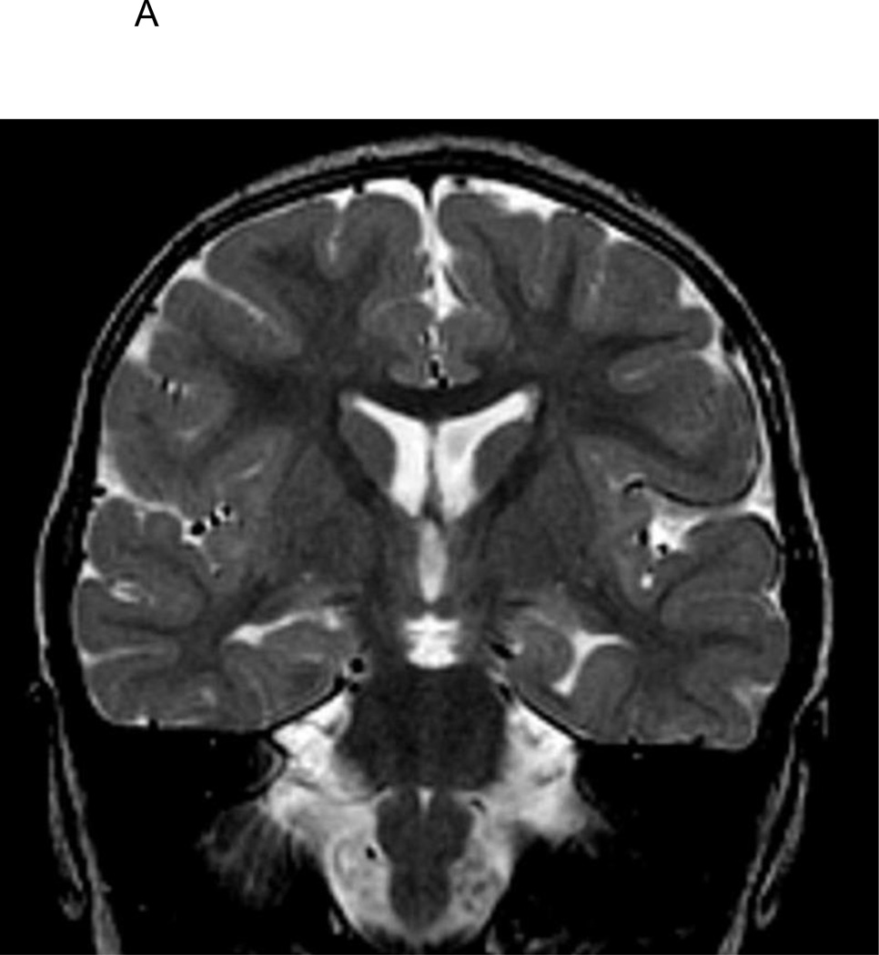
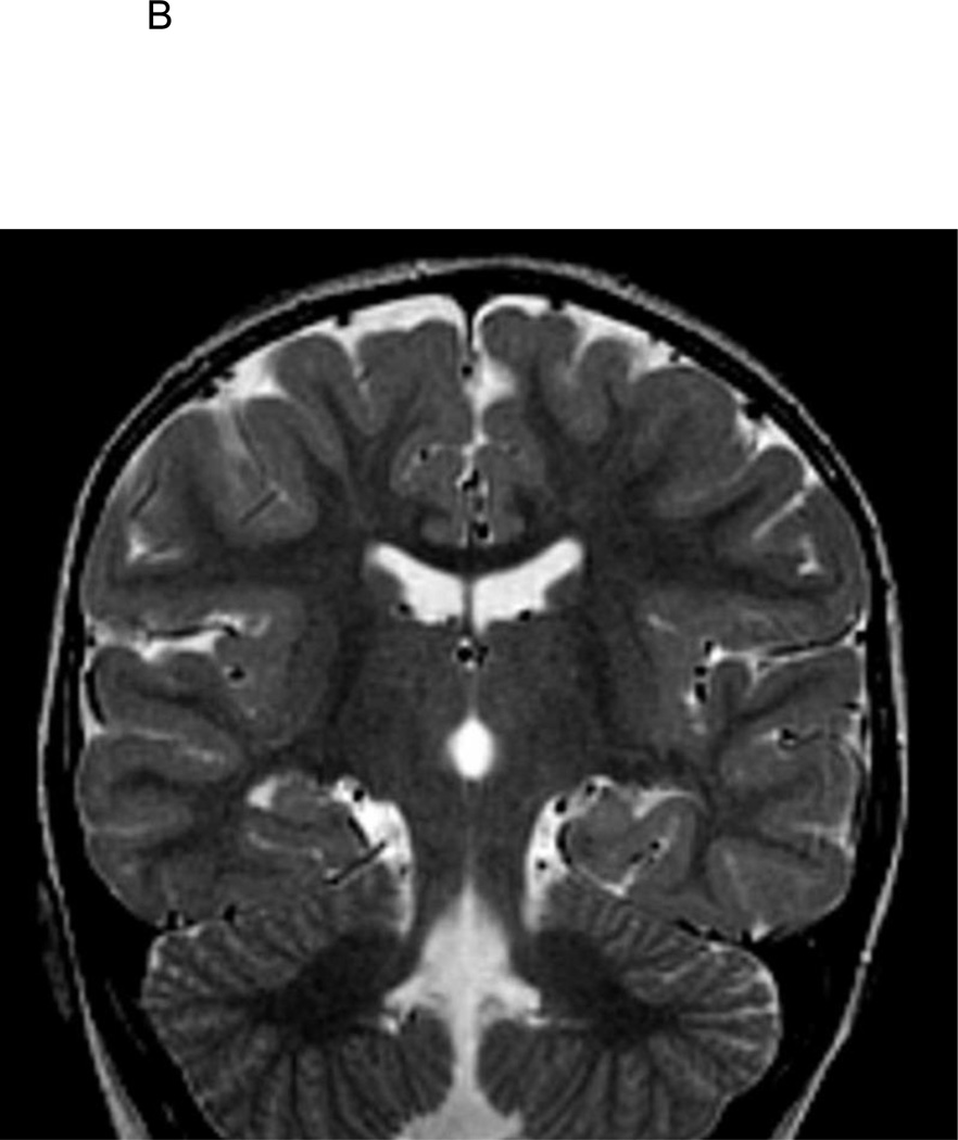
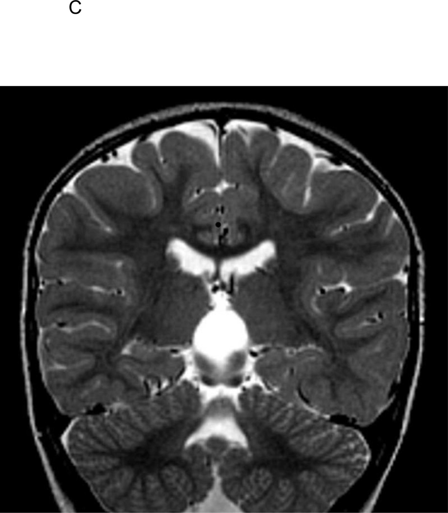
Objective: Hippocampal malrotation is characterized by incomplete hippocampal inversion with a rounded shape and blurred internal architecture. There is still debate about whether hippocampal malrotation has pathologic significance. We present findings from the Consequences of Prolonged Febrile Seizures in Childhood (FEBSTAT) study on the frequency of and risk factors for hippocampal malrotation.

Subjects and methods: FEBSTAT is a prospective multicenter study investigating the consequences of febrile status epilepticus in childhood. MRI studies of 226 patients with febrile status epilepticus were analyzed visually by two board-certified neuroradiologists blinded to clinical details and were compared with MRI studies of 96 subjects with first simple febrile seizure. Quantitative analysis of hippocampal volume was performed by two independent observers.

Results: Hippocampal malrotation was present in 20 of 226 (8.8%) patients with febrile status epilepticus compared with two of 96 (2.1%) control subjects (odds ratio [OR], 4.56; 95% CI, 1.05-19.92). Hippocampal malrotation was exclusively left-sided in 18 of 22 (81.8%) patients and bilateral in the remaining four patients (18.2%). There was no case of exclusively right-sided hippocampal malrotation. Hippocampal malrotation was more common in boys than in girls (OR, 6.1; 95% CI, 1.7-21.5). On quantitative volumetric MRI analysis, the left hippocampal volume was smaller in patients with hippocampal malrotation than in control subjects with simple febrile seizure (p = 0.004), and the right-to-left hippocampal volume ratio was higher in the hippocampal malrotation group than in the simple febrile seizure group (p < 0.001).

Conclusion: Hippocampal malrotation is a developmental malformation that predominantly affects the left hippocampus in male patients and is more frequently found in children with prolonged febrile status epilepticus than in control subjects. These data provide further evidence that hippocampal malrotation represents a pathologic error in brain development rather than a normal variant.

Keywords: MRI; febrile seizures; hippocampal malrotation; status epilepticus.


Figures

Figure 1
Figure 1
A. T2-weighted coronal image of 2 1/2-year old FSE patient demonstrating medial positioning and globular shape of the left hippocampus. B. T2-weighted coronal image of 2 1/2-year old FSE patient demonstrating medial positioning and globular shape of the left hippocampus, with associated vertically orientation of the left collateral sulcus. C. T2-weighted coronal image of 2 1/2-year old FSE patient demonstrating medial positioning and globular shape of the left hippocampus, with associated inferior positioning of the posterior left crus of the fornix.
Figure 1
Figure 1
A. T2-weighted coronal image of 2 1/2-year old FSE patient demonstrating medial positioning and globular shape of the left hippocampus. B. T2-weighted coronal image of 2 1/2-year old FSE patient demonstrating medial positioning and globular shape of the left hippocampus, with associated vertically orientation of the left collateral sulcus. C. T2-weighted coronal image of 2 1/2-year old FSE patient demonstrating medial positioning and globular shape of the left hippocampus, with associated inferior positioning of the posterior left crus of the fornix.

References

    1. Barsi P, Kenez J, Solymosi D, et al. Hippocampal malrotation with normal corpus callosum: a new entity? Neuroradiology. 2000;42:339–345. - PubMed
    1. Gamss RP, Slasky SE, Bello JA, et al. Prevalence of hippocampal malrotation in a population without seizures. AJNR Am J Neuroradiol. 2009;30:1571–1573. - PMC - PubMed
    1. Andrade DM, Krings T, Chow EW, Kiehl TR, Bassett AS. Hippocampal malrotation is associated with chromosome 22q11.2 microdeletion. Can J Neurol Sci. 2013;40:652–656. - PMC - PubMed
    1. Bajic D, Kumlien E, Mattson P, et al. Incomplete hippocampal inversion – is there a relation to epilepsy? Eur Radiol. 2009;19:2544–2550. - PubMed
    1. Bajic D, Canto Moreira N, Wikström J, et al. Asymmetric development of the hippocampal region is common: a fetal MR imaging study. AJNR Am J Neuroradiol. 2012;33(3):513–518. - PMC - PubMed

Publication types