Review
. 2015 Oct 22;163(3):560-9.
doi: 10.1016/j.cell.2015.10.001. Epub 2015 Oct 22.

Mitochondrial ROS signaling in organismal homeostasis

Affiliations
Review

Mitochondrial ROS signaling in organismal homeostasis

Gerald S Shadel et al. Cell. .

Abstract

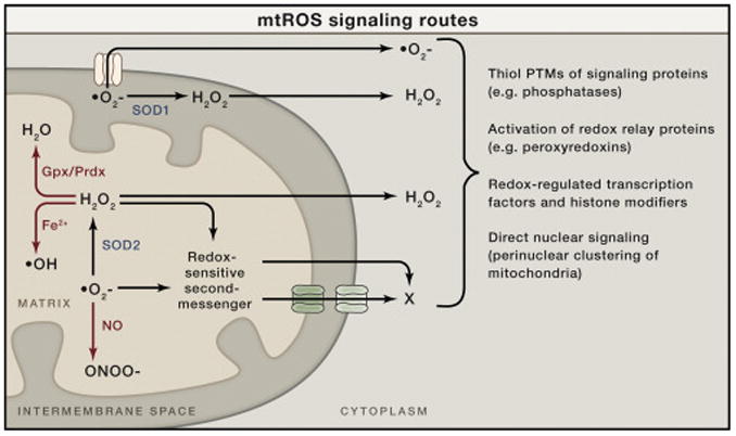
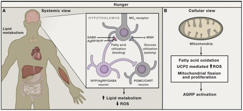
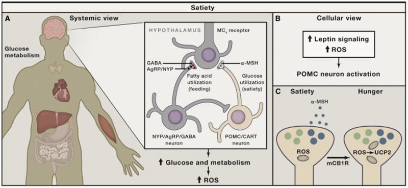
Generation, transformation, and utilization of organic molecules in support of cellular differentiation, growth, and maintenance are basic tenets that define life. In eukaryotes, mitochondrial oxygen consumption plays a central role in these processes. During the process of oxidative phosphorylation, mitochondria utilize oxygen to generate ATP from organic fuel molecules but in the process also produce reactive oxygen species (ROS). While ROS have long been appreciated for their damage-promoting, detrimental effects, there is now a greater understanding of their roles as signaling molecules. Here, we review mitochondrial ROS-mediated signaling pathways with an emphasis on how they are involved in various basal and adaptive physiological responses that control organismal homeostasis.


Figures

Figure 1
Figure 1. Mitochondrial ROS Signaling Basics
Superoxide (O2–) is generated on both sides of the inner mitochondrial membrane and hence arises in the matrix or the intermembrane space (IMS). Superoxide can be converted to hydrogen peroxide (H2O2) by superoxide dismutase enzymes (SOD1 in the IMS or SOD2 in the matrix). The resulting hydrogen peroxide can cross membranes and enter the cytoplasm to promote redox signaling. Superoxide is not readily membrane permeable but may be released into the cytoplasm through specific outer membrane channels, as shown (see main text). In addition to signaling in the cytoplasm directly, both superoxide and hydrogen peroxide could, in principle, oxidize or modify other molecules in mitochondria that can be released into the cytoplasm to signal (redox-sensitive second messenger; X). These mitochondrial ROS (mtROS) can generate signaling responses and changes in nuclear gene expression in multiple ways (shown to the right). There are other fates of mtROS that would prevent signaling (or potentially enact other signaling and damage responses). For example, superoxide can react with nitric oxide (NO) to form peroxinitrite (ONOO–). This would prevent its conversion to hydrogen peroxide, could cause damage by the highly reactive peroxynitrite, and could potentially limit NO availability for its own type of signaling. Hydrogen peroxide can be eliminated enzymatically by glutathione peroxidase (Gpx) in the matrix or peroxiredoxins (Prdx) in the matrix and elsewhere in the cell. Peroxyredoxins can also promote redox signaling by promoting disulfide bond formation in target proteins. Finally, in the presence of transition metals, hydrogen peroxide can generate damaging hydroxyl radicals (OH).
Figure 2
Figure 2. Schematic Illustration of Hypothalamic Control of Negative Energy Metabolism with Low ROS
(A) In the brain, the hypothalamus contains neuronal populations that control hunger (negative energy balance) and satiety (positive energy balance). Hunger state is promoted by neurons (green) that produce Agouti-related peptide (AgRP) and neuropeptide Y (NPY), as well as GABA. When these neurons are active (hunger, calorie restriction, starvation), systemic metabolism is shifting to lipid metabolism with an overall lower level of mtROS production in all tissues. (B) The activation of AgRP neurons during negative energy balance is promoted by pathways enabling long-chain fatty acid oxidation in the mitochondria, which is enabled by maintenance of low mtROS generation by engagement of UCP2 and mechanisms that propagate fission and/or proliferation of mitochondria (NRF1, Sirt1, and PGC1α). (C) UCP2 is believed to function as a conditional mitochondrial uncoupler in the presence of long-chain fatty acids and, as such, reduces mtROS production and overall cellular ROS levels.
Figure 3
Figure 3. Schematic Illustration of Hypothalamic Control of Positive Energy Metabolism with Elevated ROS
(A) Satiety (feeling full) is promoted by hypothalamic neurons (blue) that produce pro-opiomelanocortin (POMC)-derived peptides, such as α-MSH, which, in turn, act on melanocortin-4-receptor-containing neurons. When these neurons are active, systemic metabolism is shifting toward glucose utilization, with enhanced mtROS production contributing to increased cellular ROS in various tissues. (B) The activation of POMC neurons after a meal is accomplished by ROS, in part driven by increased mtROS production, and is supported by intracellular leptin (Jak/Stat) and insulin (PI-3K/PTEN) signaling, involving altered K-ATP channel activity. (C) Recent studies indicate that, under unique circumstances (e.g., activation of cannabinoid receptors), POMC neurons, while still driven by ROS, will become promoters of hunger rather than satiety because they shift to releasing appetite-stimulating opiates (β-endorphin) though UCP2-dependent mitochondrial adaptations.

References

    1. Abizaid A, Liu ZW, Andrews ZB, Shanabrough M, Borok E, Elsworth JD, Roth RH, Sleeman MW, Picciotto MR, Tschöp MH, et al. Ghrelin modulates the activity and synaptic input organization of midbrain dopamine neurons while promoting appetite. J Clin Invest. 2006;116:3229–3239. PubMed. - PMC - PubMed
    1. Al-Mehdi AB, Pastukh VM, Swiger BM, Reed DJ, Patel MR, Bardwell GC, Pastukh VV, Alexeyev MF, Gillespie MN. Perinuclear mitochondrial clustering creates an oxidant-rich nuclear domain required for hypoxia-induced transcription. Sci Signal. 2012;5:ra47. PubMed. - PMC - PubMed
    1. Andrews ZB, Erion D, Beiler R, Liu ZW, Abizaid A, Zigman J, Elsworth JD, Savitt JM, DiMarchi R, Tschoep M, et al. Ghrelin promotes and protects nigrostriatal dopamine function via a UCP2-dependent mitochondrial mechanism. J Neurosci. 2009;29:14057–14065. PubMed. - PMC - PubMed
    1. Andrews ZB, Horvath B, Barnstable CJ, Elsworth J, Yang L, Beal MF, Roth RH, Matthews RT, Horvath TL. Uncoupling protein-2 is critical for nigral dopamine cell survival in a mouse model of Parkinson's disease. J Neurosci. 2005;25:184–191. PubMed. - PMC - PubMed
    1. Andrews ZB, Horvath TL. Uncoupling protein-2 regulates lifespan in mice. Am J Physiol Endocrinol Metab. 2009;296:E621–E627. PubMed. - PMC - PubMed

Substances

LinkOut - more resources